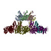
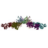
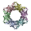
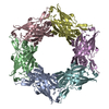
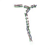
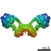
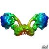
ジャーナル: J Mol Biol / 年: 2013 タイトル: The molecular architecture of the bacteriophage T4 neck. 著者: Andrei Fokine / Zhihong Zhang / Shuji Kanamaru / Valorie D Bowman / Anastasia A Aksyuk / Fumio Arisaka / Venigalla B Rao / Michael G Rossmann / 要旨: A hexamer of the bacteriophage T4 tail terminator protein, gp15, attaches to the top of the phage tail stabilizing the contractile sheath and forming the interface for binding of the independently ...A hexamer of the bacteriophage T4 tail terminator protein, gp15, attaches to the top of the phage tail stabilizing the contractile sheath and forming the interface for binding of the independently assembled head. Here we report the crystal structure of the gp15 hexamer, describe its interactions in T4 virions that have either an extended tail or a contracted tail, and discuss its structural relationship to other phage proteins. The neck of T4 virions is decorated by the "collar" and "whiskers", made of fibritin molecules. Fibritin acts as a chaperone helping to attach the long tail fibers to the virus during the assembly process. The collar and whiskers are environment-sensing devices, regulating the retraction of the long tail fibers under unfavorable conditions, thus preventing infection. Cryo-electron microscopy analysis suggests that twelve fibritin molecules attach to the phage neck with six molecules forming the collar and six molecules forming the whiskers.
履歴
登録
2012年11月12日
登録サイト: RCSB / 処理サイト: RCSB
改定 1.0
2013年3月6日
Provider: repository / タイプ: Initial release
改定 1.1
2013年5月15日
Group: Database references
改定 1.2
2018年7月18日
Group: Data collection / カテゴリ: em_software / Item: _em_software.image_processing_id
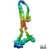
Twelve fibritin molecules attach to the bacteriophage T4 neck with six molecules forming the phage collar and six molecules forming the whiskers. The model of fibritin comprises the crystal structure of the N-terminal comain (Boudko et al., 2004, J. Mol. Biol. 339, 927-935), the crystal structure of the C-terminal domain (Tao et al., 1997, Structure 5, 789-798), and the central region, modeled with segments of triple-helical coiled coil (Letarov et al., 2005, J. Bacteriol. 187, 1055-1066).
緩衝液
名称: 50 mM Tris-HCl, pH 8.0, 0.2 M NaCl, 8 mM MgCl2 / pH: 8 / 詳細: 50 mM Tris-HCl, pH 8.0, 0.2 M NaCl, 8 mM MgCl2
モード: BRIGHT FIELD / 倍率(公称値): 38000 X / 倍率(補正後): 39190 X / 最大 デフォーカス(公称値): 3000 nm / 最小 デフォーカス(公称値): 1500 nm / Cs: 2 mm
試料ホルダ
試料ホルダーモデル: GATAN LIQUID NITROGEN
撮影
電子線照射量: 16 e/Å2 / フィルム・検出器のモデル: KODAK SO-163 FILM / 詳細: Kodak film
画像スキャン
デジタル画像の数: 97
放射
プロトコル: SINGLE WAVELENGTH / 単色(M)・ラウエ(L): M / 散乱光タイプ: x-ray
放射波長
相対比: 1
-
解析
EMソフトウェア
ID
名称
カテゴリ
1
UCSF Chimera
モデルフィッティング
2
EMAN
3次元再構成
CTF補正
詳細: phase flipping
対称性
点対称性: C1 (非対称)
3次元再構成
手法: projection matching / 解像度: 25 Å / 解像度の算出法: FSC 0.5 CUT-OFF / 粒子像の数: 2727 / ピクセルサイズ(公称値): 3.572 Å / ピクセルサイズ(実測値): 3.572 Å / 対称性のタイプ: POINT
原子モデル構築
空間: REAL 詳細: DETAILS--The model of fibritin comprises the crystal structure of the N-terminal domain (Boudko et al., 2004, J. Mol. Biol. 339, 927-935), the crystal structure of the C-terminal domain (Tao ...詳細: DETAILS--The model of fibritin comprises the crystal structure of the N-terminal domain (Boudko et al., 2004, J. Mol. Biol. 339, 927-935), the crystal structure of the C-terminal domain (Tao et al., 1997, Structure 5, 789-798), and the central region, modeled with segments of triple-helical coiled coil (Letarov et al., 2005, J. Bacteriol. 187, 1055-1066). These parts of the fibritin structure were fitted into the cryo-EM density using the program CHIMERA (Pettersen et al., 2004, J. Computat. Chem. 25, 1605-1612). Chains A, B, and C correspond to one fibritin molecule located in the phage collar. Chains D, E, and F correspond to one fibritin molecule forming the whisker.
 ムービー
ムービー コントローラー


 データを開く
 基本情報
基本情報 要素
要素 キーワード
キーワード 機能・相同性情報
 Enterobacteria phage T4 (ファージ)
 データ登録者
 引用
引用
 構造の表示
構造の表示 ダウンロードとリンク
 その他のダウンロード


 PDBj
PDBj

 集合体
集合体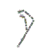

 試料調製
試料調製 電子顕微鏡撮影
 FIELD EMISSION GUN / 加速電圧: 200 kV / 照射モード: SPOT SCAN
 解析
解析