 Movie
Movie Controller


+ Open data

- Basic information
| Entry | Database: PDB / ID: 3j23 | ||||||
|---|---|---|---|---|---|---|---|
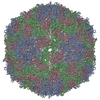
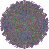
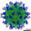
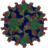
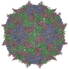
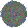
| Title | The Enterovirus 71 empty capsid | ||||||
 Components Components |
| ||||||
 Keywords Keywords | VIRUS / EV71 / 80S | ||||||
| Function / homology |  Function and homology information Function and homology informationsymbiont-mediated suppression of host cytoplasmic pattern recognition receptor signaling pathway via inhibition of MDA-5 activity / picornain 2A / symbiont-mediated suppression of host mRNA export from nucleus / symbiont genome entry into host cell via pore formation in plasma membrane / picornain 3C / T=pseudo3 icosahedral viral capsid / host cell cytoplasmic vesicle membrane / nucleoside-triphosphate phosphatase / channel activity / monoatomic ion transmembrane transport ...symbiont-mediated suppression of host cytoplasmic pattern recognition receptor signaling pathway via inhibition of MDA-5 activity / picornain 2A / symbiont-mediated suppression of host mRNA export from nucleus / symbiont genome entry into host cell via pore formation in plasma membrane / picornain 3C / T=pseudo3 icosahedral viral capsid / host cell cytoplasmic vesicle membrane / nucleoside-triphosphate phosphatase / channel activity / monoatomic ion transmembrane transport / DNA replication / RNA helicase activity / endocytosis involved in viral entry into host cell / symbiont-mediated activation of host autophagy / RNA-directed RNA polymerase / cysteine-type endopeptidase activity / viral RNA genome replication / RNA-directed RNA polymerase activity / DNA-templated transcription / virion attachment to host cell / host cell nucleus / structural molecule activity / ATP hydrolysis activity / proteolysis / RNA binding / zinc ion binding / ATP binding / membrane Similarity search - Function | ||||||
| Biological species |   Human enterovirus 71 Human enterovirus 71 | ||||||
| Method | ELECTRON MICROSCOPY / single particle reconstruction / cryo EM / Resolution: 9.2 Å | ||||||
 Authors Authors | Shingler, K.L. | ||||||
 Citation Citation | Journal: PLoS Pathog / Year: 2013 Title: The enterovirus 71 A-particle forms a gateway to allow genome release: a cryoEM study of picornavirus uncoating. Authors: Kristin L Shingler / Jennifer L Yoder / Michael S Carnegie / Robert E Ashley / Alexander M Makhov / James F Conway / Susan Hafenstein /  Abstract: Since its discovery in 1969, enterovirus 71 (EV71) has emerged as a serious worldwide health threat. This human pathogen of the picornavirus family causes hand, foot, and mouth disease, and also has ...Since its discovery in 1969, enterovirus 71 (EV71) has emerged as a serious worldwide health threat. This human pathogen of the picornavirus family causes hand, foot, and mouth disease, and also has the capacity to invade the central nervous system to cause severe disease and death. Upon binding to a host receptor on the cell surface, the virus begins a two-step uncoating process, first forming an expanded, altered "A-particle", which is primed for genome release. In a second step after endocytosis, an unknown trigger leads to RNA expulsion, generating an intact, empty capsid. Cryo-electron microscopy reconstructions of these two capsid states provide insight into the mechanics of genome release. The EV71 A-particle capsid interacts with the genome near the icosahedral two-fold axis of symmetry, which opens to the external environment via a channel ∼10 Å in diameter that is lined with patches of negatively charged residues. After the EV71 genome has been released, the two-fold channel shrinks, though the overall capsid dimensions are conserved. These structural characteristics identify the two-fold channel as the site where a gateway forms and regulates the process of genome release. | ||||||
| History |
|
- Structure visualization
| Movie |
Movie viewer |
|---|---|
| Structure viewer | Molecule: MolmilJmol/JSmol |
- Downloads & links
-Download
| PDBx/mmCIF format | 3j23.cif.gz | 147.8 KB | Display | PDBx/mmCIF format |
|---|---|---|---|---|
| PDB format | pdb3j23.ent.gz | 114.6 KB | Display | PDB format |
| PDBx/mmJSON format | 3j23.json.gz | Tree view | PDBx/mmJSON format | |
| Others |  Other downloads Other downloads |
-Validation report
| Summary document | 3j23_validation.pdf.gz | 910.4 KB | Display | wwPDB validaton report |
|---|---|---|---|---|
| Full document | 3j23_full_validation.pdf.gz | 941.6 KB | Display | |
| Data in XML | 3j23_validation.xml.gz | 29.6 KB | Display | |
| Data in CIF | 3j23_validation.cif.gz | 42.6 KB | Display | |
| Arichive directory | https://data.pdbj.org/pub/pdb/validation_reports/j2/3j23ftp://data.pdbj.org/pub/pdb/validation_reports/j2/3j23 | HTTPS FTP |
-Related structure data
| Related structure data | 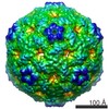 5466MC 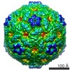 5465C  3j22C C: citing same article ( M: map data used to model this data |
|---|---|
| Similar structure data |


-Links
 PDBj
PDBj

- Assembly
| Deposited unit | 
|
|---|---|
| 1 | x 60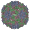
|
| 2 |
|
| 3 | x 5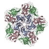
|
| 4 | x 6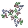
|
| 5 | 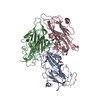
|
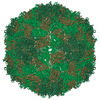
| Symmetry | Point symmetry: (Schoenflies symbol: I (icosahedral)) |
-Components
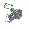
| #1: Protein | Mass: 25347.824 Da / Num. of mol.: 1 / Fragment: UNP residues 638-862 / Source method: isolated from a natural source / Source: (natural) Human enterovirus 71 / References: UniProt: B2ZUN0 |
|---|---|
| #2: Protein | Mass: 25987.320 Da / Num. of mol.: 1 / Fragment: UNP residues 82-318 / Source method: isolated from a natural source / Source: (natural) Human enterovirus 71 / References: UniProt: B2ZUN0 |
| #3: Protein | Mass: 26125.834 Da / Num. of mol.: 1 / Fragment: UNP residues 324-562 / Source method: isolated from a natural source / Source: (natural) Human enterovirus 71 / References: UniProt: B2ZUN0 |
| Sequence details | THE MODELED PROTEIN SEQUENCE WAS DERIVED FROM PDB ENTRY 3VBU. |
-Experimental details
-Experiment
| Experiment | Method: ELECTRON MICROSCOPY |
|---|---|
| EM experiment | Aggregation state: PARTICLE / 3D reconstruction method: single particle reconstruction |
- Sample preparation
| Component | Name: Human Enterovirus 71 empty capsid / Type: VIRUS Details: Icosahedral virus. The sample was purified mature virus heated in solution. |
|---|---|
| Molecular weight | Value: 7 MDa / Experimental value: NO |
| Details of virus | Empty: YES / Enveloped: NO / Host category: VERTEBRATES / Isolate: SEROTYPE / Type: VIRION |
| Natural host | Organism: Homo sapiens |
| Buffer solution | pH: 7.5 / Details: 10 mM Tris-HCL, 20 mM NaCl, 5 mM MgCl2 |
| Specimen | Conc.: 0.1 mg/ml / Embedding applied: NO / Shadowing applied: NO / Staining applied: NO / Vitrification applied: YES / Details: 10 mM Tris-HCL, 20 mM NaCl, 5 mM MgCl2 |
| Specimen support | Details: glow discharged holey carbon Quantifoil electron microscopy grids |
| Vitrification | Instrument: FEI VITROBOT MARK III / Cryogen name: OTHER / Temp: 77 K / Humidity: 95 % / Details: ETHANE-PROPANE MIXTURE |
- Electron microscopy imaging
| Experimental equipment |  Model: Tecnai F20 / Image courtesy: FEI Company |
|---|---|
| Microscopy | Model: FEI TECNAI F20 / Date: Apr 17, 2012 |
| Electron gun | Electron source:  FIELD EMISSION GUN / Accelerating voltage: 200 kV / Illumination mode: FLOOD BEAM FIELD EMISSION GUN / Accelerating voltage: 200 kV / Illumination mode: FLOOD BEAM |
| Electron lens | Mode: BRIGHT FIELD / Nominal magnification: 50000 X / Calibrated magnification: 50000 X / Nominal defocus max: 4370 nm / Nominal defocus min: 1730 nm / Cs: 2 mm / Astigmatism: CTFFIND3 (EMAN) / Camera length: 0 mm |
| Specimen holder | Specimen holder model: GATAN LIQUID NITROGEN / Temperature: 95 K / Tilt angle max: 0 ° / Tilt angle min: 0 ° |
| Image recording | Electron dose: 15 e/Å2 / Film or detector model: KODAK SO-163 FILM |
| Image scans | Num. digital images: 40 |
| Radiation | Protocol: SINGLE WAVELENGTH / Monochromatic (M) / Laue (L): M / Scattering type: x-ray |
| Radiation wavelength | Relative weight: 1 |
- Processing
| EM software |
| ||||||||||||||||||||
|---|---|---|---|---|---|---|---|---|---|---|---|---|---|---|---|---|---|---|---|---|---|
| CTF correction | Details: auto3DEM | ||||||||||||||||||||
| Symmetry | Point symmetry: I (icosahedral) | ||||||||||||||||||||
| 3D reconstruction | Method: Common lines / Resolution: 9.2 Å / Resolution method: FSC 0.5 CUT-OFF / Num. of particles: 1931 / Nominal pixel size: 1.27 Å / Actual pixel size: 1.27 Å Details: A single data set was used for the reconstruction. Particles were selected individually. Symmetry type: POINT | ||||||||||||||||||||
| Atomic model building | Protocol: RIGID BODY FIT / Space: REAL / Target criteria: correlation coefficient Details: REFINEMENT PROTOCOL--rigid body DETAILS--The protomer was fit in Chimera, used to generate a full crystal map, then docked as a rigid body in Situs. | ||||||||||||||||||||
| Atomic model building | PDB-ID: 3VBU Accession code: 3VBU / Source name: PDB / Type: experimental model | ||||||||||||||||||||
| Refinement step | Cycle: LAST
|

