 ムービー
ムービー コントローラー


+ データを開く

- 基本情報
基本情報
| 登録情報 | データベース: PDB / ID: 3izo | ||||||
|---|---|---|---|---|---|---|---|
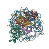
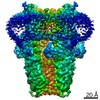
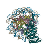
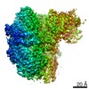
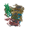
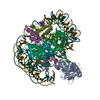
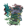
| タイトル | Model of the fiber tail and its interactions with the penton base of human adenovirus by cryo-electron microscopy | ||||||
 要素 要素 |
| ||||||
 キーワード キーワード | VIRAL PROTEIN / human adenovirus fiber tail / pentameric penton base / trimeric fiber | ||||||
| 機能・相同性 |  機能・相同性情報 機能・相同性情報T=25 icosahedral viral capsid / adhesion receptor-mediated virion attachment to host cell / viral capsid / clathrin-dependent endocytosis of virus by host cell / cell adhesion / symbiont entry into host cell / host cell nucleus / virion attachment to host cell / structural molecule activity 類似検索 - 分子機能 | ||||||
| 生物種 |   Human adenovirus 5 (ヒトアデノウイルス) Human adenovirus 5 (ヒトアデノウイルス) | ||||||
| 手法 | 電子顕微鏡法 / 単粒子再構成法 / クライオ電子顕微鏡法 / 解像度: 3.6 Å | ||||||
 データ登録者 データ登録者 | Liu, H. | ||||||
 引用 引用 | ジャーナル: J Mol Biol / 年: 2011 タイトル: Model of the trimeric fiber and its interactions with the pentameric penton base of human adenovirus by cryo-electron microscopy. 著者: Hongrong Liu / Lily Wu / Z Hong Zhou /  要旨: Adenovirus invades host cells by first binding to host receptors through a trimeric fiber, which contains three domains: a receptor-binding knob domain, a long flexible shaft domain, and a penton ...Adenovirus invades host cells by first binding to host receptors through a trimeric fiber, which contains three domains: a receptor-binding knob domain, a long flexible shaft domain, and a penton base-attachment tail domain. Although the structure of the knob domain associated with a portion of the shaft has been solved by X-ray crystallography, the in situ structure of the fiber in the virion is not known; thus, it remains a mystery how the trimeric fiber attaches to its underlying pentameric penton base. By high-resolution cryo-electron microscopy, we have determined the structure of the human adenovirus type 5 (Ad5) to 3.6-Å resolution and have reported the full atomic models for its capsid proteins, but not for the fiber whose density cannot be directly interpreted due to symmetry mismatch with the penton base. Here, we report the determination of the Ad5 fiber structure and its mode of attachment to the pentameric penton base by using an integrative approach of multi-resolution filtering, homology modeling, computational simulation of mismatched symmetries, and fitting of atomic models into cryo-electron microscopy density maps. Our structure reveals that the interactions between the trimeric fiber and the pentameric penton base are mediated by a hydrophobic ring on the top surface of the penton base and three flexible tails inserted into three of the five available grooves formed by neighboring subunits of penton base. These interaction sites provide the molecular basis for the symmetry mismatch and can be targeted for optimizing adenovirus for gene therapy applications. | ||||||
| 履歴 |
|
- 構造の表示
構造の表示
| ムービー |
ムービービューア |
|---|---|
| 構造ビューア | 分子: MolmilJmol/JSmol |
- ダウンロードとリンク
-ダウンロード
| PDBx/mmCIF形式 | 3izo.cif.gz | 440.2 KB | 表示 | PDBx/mmCIF形式 |
|---|---|---|---|---|
| PDB形式 | pdb3izo.ent.gz | 327.6 KB | 表示 | PDB形式 |
| PDBx/mmJSON形式 | 3izo.json.gz | ツリー表示 | PDBx/mmJSON形式 | |
| その他 |  その他のダウンロード その他のダウンロード |
-検証レポート
| 文書・要旨 | 3izo_validation.pdf.gz | 1 MB | 表示 | wwPDB検証レポート |
|---|---|---|---|---|
| 文書・詳細版 | 3izo_full_validation.pdf.gz | 1.2 MB | 表示 | |
| XML形式データ | 3izo_validation.xml.gz | 102.7 KB | 表示 | |
| CIF形式データ | 3izo_validation.cif.gz | 144.6 KB | 表示 | |
| アーカイブディレクトリ | https://data.pdbj.org/pub/pdb/validation_reports/iz/3izoftp://data.pdbj.org/pub/pdb/validation_reports/iz/3izo | HTTPS FTP |
-関連構造データ
| 関連構造データ | 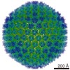 7034M M: このデータのモデリングに利用したマップデータ |
|---|---|
| 類似構造データ |


-リンク
 PDBj
PDBj

- 集合体
集合体
| 登録構造単位 | 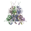
|
|---|---|
| 1 |
|
-要素
| #1: タンパク質 | 分子量: 63356.602 Da / 分子数: 5 / 由来タイプ: 天然 由来: (天然) Human adenovirus 5 (ヒトアデノウイルス)株: HEK 293 cell / 参照: UniProt: P12538 #2: タンパク質 | 分子量: 61633.066 Da / 分子数: 3 / 由来タイプ: 天然 由来: (天然) Human adenovirus 5 (ヒトアデノウイルス)株: HEK 293 cell / 参照: UniProt: Q7T416 |
|---|
-実験情報
-実験
| 実験 | 手法: 電子顕微鏡法 |
|---|---|
| EM実験 | 試料の集合状態: PARTICLE / 3次元再構成法: 単粒子再構成法 |
- 試料調製
試料調製
| 構成要素 | 名称: Human adenovirus type 5 / タイプ: VIRUS |
|---|---|
| 緩衝液 | pH: 7.5 / 詳細: 10mM Tris-HCL,1mM MgCl2 |
| 試料 | 包埋: NO / シャドウイング: NO / 染色: NO / 凍結: YES |
| 急速凍結 | 装置: FEI VITROBOT MARK I / 凍結剤: ETHANE |
- 電子顕微鏡撮影
| 実験機器 |  モデル: Titan Krios / 画像提供: FEI Company |
|---|---|
| 顕微鏡 | モデル: FEI TITAN KRIOS / 日付: 2009年5月1日 |
| 電子銃 | 電子線源:  FIELD EMISSION GUN / 加速電圧: 200 kV / 照射モード: FLOOD BEAM FIELD EMISSION GUN / 加速電圧: 200 kV / 照射モード: FLOOD BEAM |
| 電子レンズ | モード: BRIGHT FIELD / 倍率(公称値): 59000 X / 最大 デフォーカス(公称値): 2500 nm / 最小 デフォーカス(公称値): 1000 nm |
| 撮影 | 電子線照射量: 20 e/Å2 / フィルム・検出器のモデル: KODAK SO-163 FILM |
| 放射 | プロトコル: SINGLE WAVELENGTH / 単色(M)・ラウエ(L): M |
| 放射波長 | 相対比: 1 |
- 解析
解析
| EMソフトウェア | 名称: IMIRS / カテゴリ: 3次元再構成 | ||||||||||||
|---|---|---|---|---|---|---|---|---|---|---|---|---|---|
| 対称性 | 点対称性: I (正20面体型対称) | ||||||||||||
| 3次元再構成 | 手法: Cross-common lines for orientation and center refinement icosahedral symmetry-adapted functions for 3D reconstruction 解像度: 3.6 Å / 粒子像の数: 31815 / ピクセルサイズ(公称値): 1.076 Å / ピクセルサイズ(実測値): 1.1 Å / 対称性のタイプ: POINT | ||||||||||||
| 精密化ステップ | サイクル: LAST
|
