 Movie
Movie Controller


+ Open data

- Basic information
| Entry | Database: PDB / ID: 3wl7 | ||||||
|---|---|---|---|---|---|---|---|
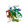
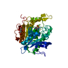
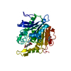
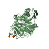
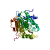
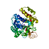
| Title | The complex structure of pOPH S172C with ligand, ACA | ||||||
 Components Components | Oxidized polyvinyl alcohol hydrolase | ||||||
 Keywords Keywords | HYDROLASE / alpha/beta-hydrolase / oxi-polyvinyl alcohol hydrolase | ||||||
| Function / homology |  Function and homology information Function and homology information | ||||||
| Biological species |  Pseudomonas sp. (bacteria) Pseudomonas sp. (bacteria) | ||||||
| Method |  X-RAY DIFFRACTION / SYNCHROTRON / MOLECULAR REPLACEMENT / Resolution: 1.67 Å X-RAY DIFFRACTION / SYNCHROTRON / MOLECULAR REPLACEMENT / Resolution: 1.67 Å | ||||||
 Authors Authors | Yang, Y. / Ko, T.P. / Li, J.H. / Liu, L. / Huang, C.H. / Chan, H.C. / Ren, F.F. / Jia, D.X. / Wang, A.H.-J. / Guo, R.T. ...Yang, Y. / Ko, T.P. / Li, J.H. / Liu, L. / Huang, C.H. / Chan, H.C. / Ren, F.F. / Jia, D.X. / Wang, A.H.-J. / Guo, R.T. / Chen, J. / Du, G.C. | ||||||
 Citation Citation | Journal: Chembiochem / Year: 2014 Title: Structural insights into enzymatic degradation of oxidized polyvinyl alcohol Authors: Yang, Y. / Ko, T.P. / Liu, L. / Li, J. / Huang, C.H. / Chan, H.C. / Ren, F. / Jia, D. / Wang, A.H.-J. / Guo, R.T. / Chen, J. / Du, G. | ||||||
| History |
|
- Structure visualization
| Structure viewer | Molecule: MolmilJmol/JSmol |
|---|
- Downloads & links
-Download
| PDBx/mmCIF format | 3wl7.cif.gz | 87.3 KB | Display | PDBx/mmCIF format |
|---|---|---|---|---|
| PDB format | pdb3wl7.ent.gz | 63.5 KB | Display | PDB format |
| PDBx/mmJSON format | 3wl7.json.gz | Tree view | PDBx/mmJSON format | |
| Others |  Other downloads Other downloads |
-Validation report
| Arichive directory | https://data.pdbj.org/pub/pdb/validation_reports/wl/3wl7ftp://data.pdbj.org/pub/pdb/validation_reports/wl/3wl7 | HTTPS FTP |
|---|
-Related structure data
| Related structure data |  3wl5SC 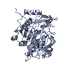 3wl6C 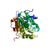 3wl8C  3wlaC S: Starting model for refinement C: citing same article ( |
|---|---|
| Similar structure data |

-Links
 PDBj
PDBj- Assembly
| Deposited unit | 
| ||||||||
|---|---|---|---|---|---|---|---|---|---|
| 1 |
| ||||||||
| Unit cell |
|
-Components
| #1: Protein | Mass: 39328.789 Da / Num. of mol.: 1 / Fragment: UNP residues 30-379 / Mutation: S172C Source method: isolated from a genetically manipulated source Source: (gene. exp.) Pseudomonas sp. (bacteria) / Strain: VM15C / Gene: pvaB / Plasmid: pET32a / Production host: |
|---|---|
| #2: Chemical | ChemComp-P2D /   Mass: 100.116 Da / Num. of mol.: 1 / Source method: obtained synthetically / Formula: C5H8O2 Mass: 100.116 Da / Num. of mol.: 1 / Source method: obtained synthetically / Formula: C5H8O2 |
| #3: Chemical | ChemComp-CIT /   Mass: 192.124 Da / Num. of mol.: 1 / Source method: obtained synthetically / Formula: C6H8O7 Mass: 192.124 Da / Num. of mol.: 1 / Source method: obtained synthetically / Formula: C6H8O7 |
| #4: Water | ChemComp-HOH /  Mass: 18.015 Da / Num. of mol.: 362 / Source method: isolated from a natural source / Formula: H2O Mass: 18.015 Da / Num. of mol.: 362 / Source method: isolated from a natural source / Formula: H2O |
-Experimental details
-Experiment
| Experiment | Method: X-RAY DIFFRACTION / Number of used crystals: 1 |
|---|
- Sample preparation
| Crystal | Density Matthews: 2.06 Å3/Da / Density % sol: 40.31 % |
|---|---|
| Crystal grow | Temperature: 295 K / Method: vapor diffusion, sitting drop / pH: 5.6 Details: 0.1M tri-sodium citrate, pH 5.6, 28% w/v Polyethylene Glycol 4000, 5% w/v n-Octyl-beta-D-glucoside, VAPOR DIFFUSION, SITTING DROP, temperature 295K |
-Data collection
| Diffraction | Mean temperature: 100 K |
|---|---|
| Diffraction source | Source: SYNCHROTRON / Site: NSRRC  / Beamline: BL13B1 / Wavelength: 1 Å / Beamline: BL13B1 / Wavelength: 1 Å |
| Detector | Type: ADSC QUANTUM 315 / Detector: CCD / Date: Jun 29, 2013 |
| Radiation | Monochromator: SI 111 CHANNEL / Protocol: SINGLE WAVELENGTH / Monochromatic (M) / Laue (L): M / Scattering type: x-ray |
| Radiation wavelength | Wavelength: 1 Å / Relative weight: 1 |
| Reflection | Resolution: 1.67→25 Å / Num. all: 38402 / Num. obs: 37918 / % possible obs: 99.3 % / Observed criterion σ(F): 0 / Observed criterion σ(I): 3 / Redundancy: 7.6 % / Rmerge(I) obs: 0.059 / Net I/σ(I): 41.7 |
| Reflection shell | Resolution: 1.67→1.73 Å / Redundancy: 6.9 % / Rmerge(I) obs: 0.19 / Mean I/σ(I) obs: 8 / Num. unique all: 3681 / % possible all: 98.1 |
- Processing
| Software |
| ||||||||||||||||||||||||||||
|---|---|---|---|---|---|---|---|---|---|---|---|---|---|---|---|---|---|---|---|---|---|---|---|---|---|---|---|---|---|
| Refinement | Method to determine structure: MOLECULAR REPLACEMENT Starting model: PDB ENTRY 3WL5 Resolution: 1.67→25 Å / Occupancy max: 1 / Occupancy min: 1 / σ(F): 0 / Stereochemistry target values: Engh & Huber
| ||||||||||||||||||||||||||||
| Solvent computation | Bsol: 34.7034 Å2 | ||||||||||||||||||||||||||||
| Displacement parameters | Biso max: 47.77 Å2 / Biso mean: 17.1829 Å2 / Biso min: 7.59 Å2
| ||||||||||||||||||||||||||||
| Refine analyze |
| ||||||||||||||||||||||||||||
| Refinement step | Cycle: LAST / Resolution: 1.67→25 Å
| ||||||||||||||||||||||||||||
| Refine LS restraints |
| ||||||||||||||||||||||||||||
| LS refinement shell | Resolution: 1.67→1.73 Å
|
