 Movie
Movie Controller


+ Open data

- Basic information
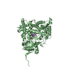
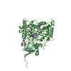
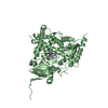
| Entry | Database: PDB / ID: 3ruk | ||||||
|---|---|---|---|---|---|---|---|
| Title | Human Cytochrome P450 CYP17A1 in complex with Abiraterone | ||||||
 Components Components | Steroid 17-alpha-hydroxylase/17,20 lyase | ||||||
 Keywords Keywords | OXIDOREDUCTASE/OXIDOREDUCTASE INHIBITOR / cytochrome P450 / CYP17A1 / P450 17A1 / monooxygenase / 17a-hydroxylase / 17 / 20-lyase / heme protein / cytochrome P450 oxidoreductase / Abiraterone / Zytiga / 17a-hydroxylation / membrane / microsome / endoplasmic reticulum / Galeterone / OXIDOREDUCTASE-OXIDOREDUCTASE INHIBITOR complex | ||||||
| Function / homology |  Function and homology information Function and homology informationDefective CYP17A1 causes AH5 / steroid 17alpha-monooxygenase / 17alpha-hydroxyprogesterone deacetylase / steroid 17-alpha-monooxygenase activity / cortisol biosynthetic process / Androgen biosynthesis / glucocorticoid biosynthetic process / hormone biosynthetic process / Glucocorticoid biosynthesis / androgen biosynthetic process ...Defective CYP17A1 causes AH5 / steroid 17alpha-monooxygenase / 17alpha-hydroxyprogesterone deacetylase / steroid 17-alpha-monooxygenase activity / cortisol biosynthetic process / Androgen biosynthesis / glucocorticoid biosynthetic process / hormone biosynthetic process / Glucocorticoid biosynthesis / androgen biosynthetic process / sex differentiation / progesterone metabolic process / steroid biosynthetic process / steroid metabolic process / oxygen binding / lyase activity / iron ion binding / axon / neuronal cell body / heme binding / endoplasmic reticulum membrane / endoplasmic reticulum Similarity search - Function | ||||||
| Biological species |  Homo sapiens (human) Homo sapiens (human) | ||||||
| Method |  X-RAY DIFFRACTION / SYNCHROTRON / MOLECULAR REPLACEMENT / Resolution: 2.6 Å X-RAY DIFFRACTION / SYNCHROTRON / MOLECULAR REPLACEMENT / Resolution: 2.6 Å | ||||||
 Authors Authors | DeVore, N.M. / Scott, E.E. | ||||||
 Citation Citation | Journal: Nature / Year: 2012 Title: Structures of cytochrome P450 17A1 with prostate cancer drugs abiraterone and TOK-001. Authors: Devore, N.M. / Scott, E.E. | ||||||
| History |
|
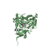
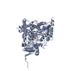
- Structure visualization
| Structure viewer | Molecule: MolmilJmol/JSmol |
|---|
- Downloads & links
-Download
| PDBx/mmCIF format | 3ruk.cif.gz | 378.5 KB | Display | PDBx/mmCIF format |
|---|---|---|---|---|
| PDB format | pdb3ruk.ent.gz | 309.6 KB | Display | PDB format |
| PDBx/mmJSON format | 3ruk.json.gz | Tree view | PDBx/mmJSON format | |
| Others |  Other downloads Other downloads |
-Validation report
| Arichive directory | https://data.pdbj.org/pub/pdb/validation_reports/ru/3rukftp://data.pdbj.org/pub/pdb/validation_reports/ru/3ruk | HTTPS FTP |
|---|
-Related structure data


-Links
 PDBj
PDBj

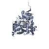
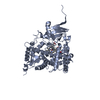
- Assembly
| Deposited unit | 
| ||||||||
|---|---|---|---|---|---|---|---|---|---|
| 1 | 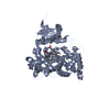
| ||||||||
| 2 | 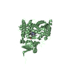
| ||||||||
| 3 | 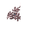
| ||||||||
| 4 | 
| ||||||||
| Unit cell |
|
-Components
| #1: Protein | Mass: 55740.141 Da / Num. of mol.: 4 / Fragment: unp residues 24-508 Source method: isolated from a genetically manipulated source Source: (gene. exp.) Homo sapiens (human) / Gene: CYP17, CYP17A1, S17AH / Plasmid: pCWori+ / Production host:  #2: Chemical | ChemComp-HEM /   Mass: 616.487 Da / Num. of mol.: 4 / Source method: obtained synthetically / Formula: C34H32FeN4O4 Mass: 616.487 Da / Num. of mol.: 4 / Source method: obtained synthetically / Formula: C34H32FeN4O4#3: Chemical | ChemComp-AER / 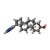  Mass: 349.509 Da / Num. of mol.: 4 / Source method: obtained synthetically / Formula: C24H31NO Mass: 349.509 Da / Num. of mol.: 4 / Source method: obtained synthetically / Formula: C24H31NO#4: Water | ChemComp-HOH / |  Mass: 18.015 Da / Num. of mol.: 118 / Source method: isolated from a natural source / Formula: H2O Mass: 18.015 Da / Num. of mol.: 118 / Source method: isolated from a natural source / Formula: H2O |
|---|
-Experimental details
-Experiment
| Experiment | Method: X-RAY DIFFRACTION / Number of used crystals: 1 |
|---|
- Sample preparation
| Crystal | Density Matthews: 2.53 Å3/Da / Density % sol: 51.38 % |
|---|---|
| Crystal grow | Temperature: 298 K / pH: 8.5 Details: 30% PEG 3350, 0.175 M Tris, 0.30 M ammonium sulfate, 3% glycerol, pH 8.5, VAPOR DIFFUSION, HANGING DROP, temperature 298K |
-Data collection
| Diffraction | Mean temperature: 100 K |
|---|---|
| Diffraction source | Source: SYNCHROTRON / Site: SSRL  / Beamline: BL9-2 / Wavelength: 0.97946 / Beamline: BL9-2 / Wavelength: 0.97946 |
| Detector | Type: MARMOSAIC 325 mm CCD / Detector: CCD / Date: Nov 26, 2010 / Details: RH COATED FLAT MIRROR, TOROIDAL FOCUSING MIRROR |
| Radiation | Monochromator: DOUBLE CRYSTAL MONOCHROMATOR / Protocol: SINGLE WAVELENGTH / Monochromatic (M) / Laue (L): M / Scattering type: x-ray |
| Radiation wavelength | Wavelength: 0.97946 Å / Relative weight: 1 |
| Reflection | Resolution: 2.6→40.49 Å / Num. obs: 70364 / % possible obs: 100 % / Observed criterion σ(I): 0 / Redundancy: 7.5 % / Biso Wilson estimate: 56.2 Å2 / Net I/σ(I): 9.6 |
| Reflection shell | Resolution: 2.6→2.67 Å / Redundancy: 7.5 % / Mean I/σ(I) obs: 2.1 / % possible all: 100 |
- Processing
| Software |
| |||||||||||||||||||||||||
|---|---|---|---|---|---|---|---|---|---|---|---|---|---|---|---|---|---|---|---|---|---|---|---|---|---|---|
| Refinement | Method to determine structure: MOLECULAR REPLACEMENT Starting model: CYP2R1 Resolution: 2.6→40.49 Å / Cor.coef. Fo:Fc: 0.938 / Cor.coef. Fo:Fc free: 0.89 / SU B: 13.297 / SU ML: 0.28 / Cross valid method: THROUGHOUT / σ(F): 0 / ESU R Free: 0.368 Stereochemistry target values: MAXIMUM LIKELIHOOD WITH PHASES
| |||||||||||||||||||||||||
| Solvent computation | Ion probe radii: 0.8 Å / Shrinkage radii: 0.8 Å / VDW probe radii: 1.4 Å / Solvent model: MASK | |||||||||||||||||||||||||
| Displacement parameters | Biso mean: 42.64 Å2
| |||||||||||||||||||||||||
| Refine analyze | Luzzati coordinate error obs: 0.3442 Å | |||||||||||||||||||||||||
| Refinement step | Cycle: LAST / Resolution: 2.6→40.49 Å
| |||||||||||||||||||||||||
| LS refinement shell | Resolution: 2.6→2.667 Å / Total num. of bins used: 20
|
