Protocol: SINGLE WAVELENGTH / Monochromatic (M) / Laue (L): M / Scattering type: x-ray
Radiation wavelength
Wavelength: 0.97924 Å / Relative weight: 1
Reflection
Redundancy: 3.9 % / Av σ(I) over netI: 5.4 / Number: 233131 / Rsym value: 0.091 / D res high: 2.09 Å / D res low: 36.263 Å / Num. obs: 59553 / % possible obs: 98.2
Diffraction reflection shell
Highest resolution (Å)
Lowest resolution (Å)
% possible obs (%)
ID
Rmerge(I) obs
Rsym value
Redundancy
6.61
36.26
98.3
1
0.04
0.04
3.8
4.67
6.61
99.3
1
0.062
0.062
3.9
3.82
4.67
99.1
1
0.063
0.063
3.9
3.3
3.82
99.1
1
0.066
0.066
3.9
2.96
3.3
98.8
1
0.089
0.089
3.9
2.7
2.96
98.4
1
0.127
0.127
3.9
2.5
2.7
97.9
1
0.191
0.191
3.9
2.34
2.5
97.8
1
0.299
0.299
3.9
2.2
2.34
97.9
1
0.468
0.468
3.9
2.09
2.2
97.1
1
0.755
0.755
3.9
Reflection
Resolution: 2.09→36.263 Å / Num. all: 60645 / Num. obs: 59553 / % possible obs: 98.2 % / Redundancy: 3.9 % / Biso Wilson estimate: 36.3 Å2 / Rmerge(I) obs: 0.091 / Rsym value: 0.091 / Net I/σ(I): 8.4
Reflection shell
Resolution (Å)
Redundancy (%)
Rmerge(I) obs
Mean I/σ(I) obs
Num. measured all
Num. unique all
Rsym value
% possible all
2.09-2.2
3.9
0.755
1
33838
8602
0.755
97.1
2.2-2.34
3.9
0.468
1.6
31911
8123
0.468
97.9
2.34-2.5
3.9
0.299
2.5
30386
7719
0.299
97.8
2.5-2.7
3.9
0.191
3.9
28066
7140
0.191
97.9
2.7-2.96
3.9
0.127
5.5
26053
6638
0.127
98.4
2.96-3.3
3.9
0.089
7.4
23595
6023
0.089
98.8
3.3-3.82
3.9
0.066
8.9
20835
5338
0.066
99.1
3.82-4.67
3.9
0.063
8.9
17533
4515
0.063
99.1
4.67-6.61
3.9
0.062
7.9
13574
3516
0.062
99.3
6.61-36.263
3.8
0.04
13.5
7340
1939
0.04
98.3
-
Phasing
Phasing
Method: molecular replacement
Phasing MR
Rfactor: 40.13 / Model details: Phaser MODE: MR_AUTO
Highest resolution
Lowest resolution
Rotation
2.5 Å
36.26 Å
Translation
2.5 Å
36.26 Å
-
Processing
Software
Name
Version
Classification
NB
SCALA
3.3.9
datascaling
PHASER
2.1.4
phasing
REFMAC
refinement
PDB_EXTRACT
3.1
dataextraction
CrystalClear
datacollection
MOSFLM
datareduction
Refinement
Method to determine structure: MOLECULAR REPLACEMENT Starting model: Ensemble of 2EXE, 2EU9, 1Z57
Resolution: 2.09→36.263 Å / Cor.coef. Fo:Fc: 0.96 / Cor.coef. Fo:Fc free: 0.945 / WRfactor Rfree: 0.2287 / WRfactor Rwork: 0.1875 / Occupancy max: 1 / Occupancy min: 0.5 / FOM work R set: 0.87 / SU B: 8.663 / SU ML: 0.117 / SU R Cruickshank DPI: 0.1611 / SU Rfree: 0.1498 / Cross valid method: THROUGHOUT / σ(F): 0 / ESU R Free: 0.15 / Stereochemistry target values: MAXIMUM LIKELIHOOD Details: HYDROGENS HAVE BEEN ADDED IN THE RIDING POSITIONS U VALUES: WITH TLS ADDED
Rfactor
Num. reflection
% reflection
Selection details
Rfree
0.2193
1988
3.3 %
RANDOM
Rwork
0.1809
-
-
-
all
0.1822
60691
-
-
obs
0.1822
59520
98.07 %
-
Solvent computation
Ion probe radii: 0.8 Å / Shrinkage radii: 0.8 Å / VDW probe radii: 1.4 Å / Solvent model: MASK
In the structure databanks used in Yorodumi, some data are registered as the other names, "COVID-19 virus" and "2019-nCoV". Here are the details of the virus and the list of structure data.
Jan 31, 2019. EMDB accession codes are about to change! (news from PDBe EMDB page)
EMDB accession codes are about to change! (news from PDBe EMDB page)
The allocation of 4 digits for EMDB accession codes will soon come to an end. Whilst these codes will remain in use, new EMDB accession codes will include an additional digit and will expand incrementally as the available range of codes is exhausted. The current 4-digit format prefixed with “EMD-” (i.e. EMD-XXXX) will advance to a 5-digit format (i.e. EMD-XXXXX), and so on. It is currently estimated that the 4-digit codes will be depleted around Spring 2019, at which point the 5-digit format will come into force.
The EM Navigator/Yorodumi systems omit the EMD- prefix.
Related info.:Q: What is EMD? / ID/Accession-code notation in Yorodumi/EM Navigator
Yorodumi is a browser for structure data from EMDB, PDB, SASBDB, etc.
This page is also the successor to EM Navigator detail page, and also detail information page/front-end page for Omokage search.
The word "yorodu" (or yorozu) is an old Japanese word meaning "ten thousand". "mi" (miru) is to see.
Related info.:EMDB / PDB / SASBDB / Comparison of 3 databanks / Yorodumi Search / Aug 31, 2016. New EM Navigator & Yorodumi / Yorodumi Papers / Jmol/JSmol / Function and homology information / Changes in new EM Navigator and Yorodumi
 Movie
Movie Controller

 Yorodumi
 Open data
 Basic information
 Components
 Keywords
 Function and homology information
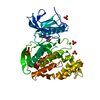
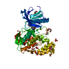
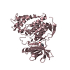
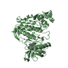
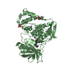
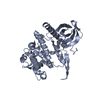
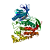
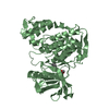
 Homo sapiens (human)
 X-RAY DIFFRACTION /
  Authors
 Citation
 Structure visualization
 Downloads & links
 Other downloads
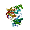


 PDBj
PDBj Assembly
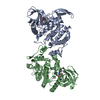


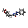
 Mass: 307.303 Da / Num. of mol.: 2 / Source method: obtained synthetically / Formula: C17H13N3O3
 Mass: 18.015 Da / Num. of mol.: 246 / Source method: isolated from a natural source / Formula: H2O
 Sample preparation
 / Beamline: X10SA / Wavelength: 0.97924 Å
 Processing