 Movie
Movie Controller

[English] 日本語
 Yorodumi
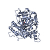
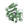
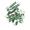
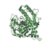
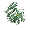
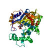
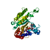
- PDB-3pax: THE CATALYTIC FRAGMENT OF POLY(ADP-RIBOSE) POLYMERASE COMPLEXED W... -
+ Open data

- Basic information
| Entry | Database: PDB / ID: 3pax | ||||||
|---|---|---|---|---|---|---|---|
| Title | THE CATALYTIC FRAGMENT OF POLY(ADP-RIBOSE) POLYMERASE COMPLEXED WITH 3-METHOXYBENZAMIDE | ||||||
 Components Components | POLY(ADP-RIBOSE) POLYMERASE | ||||||
 Keywords Keywords | TRANSFERASE / GLYCOSYLTRANSFERASE / NAD(+) ADP-RIBOSYLTRANSFERASE | ||||||
| Function / homology |  Function and homology information Function and homology informationNAD+-protein-tyrosine ADP-ribosyltransferase activity / NAD+-protein-histidine ADP-ribosyltransferase activity / NAD+-protein-serine ADP-ribosyltransferase activity / DNA ADP-ribosylation / ATP generation from poly-ADP-D-ribose / replication fork reversal / NAD+ ADP-ribosyltransferase / protein auto-ADP-ribosylation / NAD+-protein-aspartate ADP-ribosyltransferase activity / protein poly-ADP-ribosylation ...NAD+-protein-tyrosine ADP-ribosyltransferase activity / NAD+-protein-histidine ADP-ribosyltransferase activity / NAD+-protein-serine ADP-ribosyltransferase activity / DNA ADP-ribosylation / ATP generation from poly-ADP-D-ribose / replication fork reversal / NAD+ ADP-ribosyltransferase / protein auto-ADP-ribosylation / NAD+-protein-aspartate ADP-ribosyltransferase activity / protein poly-ADP-ribosylation / NAD+-protein-glutamate ADP-ribosyltransferase activity / NAD+-protein mono-ADP-ribosyltransferase activity / nuclear replication fork / Transferases; Glycosyltransferases; Pentosyltransferases / NAD+ poly-ADP-ribosyltransferase activity / nucleosome binding / positive regulation of double-strand break repair via homologous recombination / nucleotidyltransferase activity / negative regulation of innate immune response / NAD binding / double-strand break repair / site of double-strand break / damaged DNA binding / innate immune response / chromatin / nucleolus / negative regulation of transcription by RNA polymerase II / protein homodimerization activity / zinc ion binding / cytosol Similarity search - Function | ||||||
| Biological species |  | ||||||
| Method |  X-RAY DIFFRACTION / SYNCHROTRON / DIFFERENCE FOURIER / Resolution: 2.4 Å X-RAY DIFFRACTION / SYNCHROTRON / DIFFERENCE FOURIER / Resolution: 2.4 Å | ||||||
 Authors Authors | Ruf, A. / Schulz, G.E. | ||||||
 Citation Citation | Journal: Biochemistry / Year: 1998 Title: Inhibitor and NAD+ binding to poly(ADP-ribose) polymerase as derived from crystal structures and homology modeling. Authors: Ruf, A. / de Murcia, G. / Schulz, G.E. #1: Journal: Proc.Natl.Acad.Sci.USA / Year: 1996Title: Structure of the Catalytic Fragment of Poly(Ad-Ribose) Polymerase from Chicken Authors: Ruf, A. / Mennissier De Murcia, J. / De Murcia, G.M. / Schulz, G.E. #2: Journal: J.Mol.Biol. / Year: 1994Title: Crystallization and X-Ray Crystallographic Analysis of Recombinant Chicken Poly(Adp-Ribose) Polymerase Catalytic Domain Produced in Sf9 Insect Cells Authors: Jung, S. / Miranda, E.A. / De Murcia, J.M. / Niedergang, C. / Delarue, M. / Schulz, G.E. / De Murcia, G.M. | ||||||
| History |
|
- Structure visualization
| Structure viewer | Molecule: MolmilJmol/JSmol |
|---|
- Downloads & links
-Download
| PDBx/mmCIF format | 3pax.cif.gz | 82.9 KB | Display | PDBx/mmCIF format |
|---|---|---|---|---|
| PDB format | pdb3pax.ent.gz | 61.8 KB | Display | PDB format |
| PDBx/mmJSON format | 3pax.json.gz | Tree view | PDBx/mmJSON format | |
| Others |  Other downloads Other downloads |
-Validation report
| Arichive directory | https://data.pdbj.org/pub/pdb/validation_reports/pa/3paxftp://data.pdbj.org/pub/pdb/validation_reports/pa/3pax | HTTPS FTP |
|---|
-Related structure data
| Related structure data |  2pawC  2paxC  4paxC  1paxS S: Starting model for refinement C: citing same article ( |
|---|---|
| Similar structure data |


-Links
 PDBj
PDBj

- Assembly
| Deposited unit | 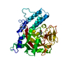
| ||||||||
|---|---|---|---|---|---|---|---|---|---|
| 1 |
| ||||||||
| Unit cell |
|
-Components
| #1: Protein | Mass: 40415.352 Da / Num. of mol.: 1 / Fragment: CATALYTIC FRAGMENT Source method: isolated from a genetically manipulated source Source: (gene. exp.)   Spodoptera frugiperda (fall armyworm) / Strain (production host): SF9 / References: UniProt: P26446, NAD+ ADP-ribosyltransferase Spodoptera frugiperda (fall armyworm) / Strain (production host): SF9 / References: UniProt: P26446, NAD+ ADP-ribosyltransferase |
|---|---|
| #2: Chemical | ChemComp-3MB / 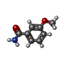  Mass: 151.163 Da / Num. of mol.: 1 / Source method: obtained synthetically / Formula: C8H9NO2 Mass: 151.163 Da / Num. of mol.: 1 / Source method: obtained synthetically / Formula: C8H9NO2 |
| #3: Water | ChemComp-HOH /  Mass: 18.015 Da / Num. of mol.: 76 / Source method: isolated from a natural source / Formula: H2O Mass: 18.015 Da / Num. of mol.: 76 / Source method: isolated from a natural source / Formula: H2O |
| Sequence details | HUMAN SEQUENCE NUMBERS ARE USED THROUGHOUT INSTEAD OF CHICKEN NUMBERS TO FACILITATE COMPARISON WITH ...HUMAN SEQUENCE NUMBERS ARE USED THROUGHOUT |
-Experimental details
-Experiment
| Experiment | Method: X-RAY DIFFRACTION / Number of used crystals: 1 |
|---|
- Sample preparation
| Crystal | Density Matthews: 2.3 Å3/Da / Density % sol: 47 % | |||||||||||||||||||||||||||||||||||
|---|---|---|---|---|---|---|---|---|---|---|---|---|---|---|---|---|---|---|---|---|---|---|---|---|---|---|---|---|---|---|---|---|---|---|---|---|
| Crystal grow | pH: 8.5 Details: PROTEIN WAS CRYSTALLIZED FROM 12% PEG 600, 6% ISOPROPANOL, 100 MM TRIS, PH 8.5, 5MM 3-METHOXYBENZAMIDE | |||||||||||||||||||||||||||||||||||
| Crystal grow | *PLUS Method: vapor diffusion, hanging drop | |||||||||||||||||||||||||||||||||||
| Components of the solutions | *PLUS
|
-Data collection
| Diffraction | Mean temperature: 293 K |
|---|---|
| Diffraction source | Source: SYNCHROTRON / Site: EMBL/DESY, HAMBURG  / Beamline: X31 / Wavelength: 0.98 / Beamline: X31 / Wavelength: 0.98 |
| Detector | Type: MAR scanner 180 mm plate / Detector: IMAGE PLATE / Date: Oct 18, 1995 |
| Radiation | Monochromatic (M) / Laue (L): M / Scattering type: x-ray |
| Radiation wavelength | Wavelength: 0.98 Å / Relative weight: 1 |
| Reflection | Resolution: 2.4→19.8 Å / Num. obs: 15091 / % possible obs: 99.5 % / Observed criterion σ(I): 0 / Redundancy: 3.7 % / Biso Wilson estimate: 37 Å2 / Rmerge(I) obs: 0.072 / Rsym value: 0.072 / Net I/σ(I): 9.7 |
| Reflection shell | Resolution: 2.4→2.42 Å / Redundancy: 3.7 % / Rmerge(I) obs: 0.394 / Mean I/σ(I) obs: 3.2 / Rsym value: 0.394 / % possible all: 100 |
| Reflection | *PLUS % possible obs: 99 % / Num. measured all: 56084 |
| Reflection shell | *PLUS % possible obs: 100 % |
- Processing
| Software |
| ||||||||||||||||||||||||||||||||||||||||||||||||||||||||||||
|---|---|---|---|---|---|---|---|---|---|---|---|---|---|---|---|---|---|---|---|---|---|---|---|---|---|---|---|---|---|---|---|---|---|---|---|---|---|---|---|---|---|---|---|---|---|---|---|---|---|---|---|---|---|---|---|---|---|---|---|---|---|
| Refinement | Method to determine structure: DIFFERENCE FOURIER Starting model: PDB ENTRY 1PAX Resolution: 2.4→19.8 Å / Isotropic thermal model: RESTRAINED / Details: X-PLOR BULK SOLVENT CORRECTION WAS APPLIED.
| ||||||||||||||||||||||||||||||||||||||||||||||||||||||||||||
| Displacement parameters | Biso mean: 35 Å2 | ||||||||||||||||||||||||||||||||||||||||||||||||||||||||||||
| Refine analyze | Luzzati d res low obs: 5 Å / Luzzati sigma a obs: 0.29 Å | ||||||||||||||||||||||||||||||||||||||||||||||||||||||||||||
| Refinement step | Cycle: LAST / Resolution: 2.4→19.8 Å
| ||||||||||||||||||||||||||||||||||||||||||||||||||||||||||||
| Refine LS restraints |
| ||||||||||||||||||||||||||||||||||||||||||||||||||||||||||||
| Xplor file |
| ||||||||||||||||||||||||||||||||||||||||||||||||||||||||||||
| Software | *PLUS Name: X-PLOR / Version: 3.851 / Classification: refinement | ||||||||||||||||||||||||||||||||||||||||||||||||||||||||||||
| Refinement | *PLUS Lowest resolution: 20 Å | ||||||||||||||||||||||||||||||||||||||||||||||||||||||||||||
| Solvent computation | *PLUS | ||||||||||||||||||||||||||||||||||||||||||||||||||||||||||||
| Displacement parameters | *PLUS |
