 Movie
Movie Controller


+ Open data

- Basic information
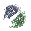
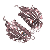
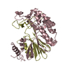
| Entry | Database: PDB / ID: 3kdh | ||||||
|---|---|---|---|---|---|---|---|
| Title | Structure of ligand-free PYL2 | ||||||
 Components Components | Putative uncharacterized protein At2g26040 | ||||||
 Keywords Keywords | HORMONE RECEPTOR / PYL2 | ||||||
| Function / homology |  Function and homology information Function and homology informationprotein phosphatase inhibitor complex / abscisic acid binding / abscisic acid-activated signaling pathway / protein phosphatase inhibitor activity / signaling receptor activity / protein homodimerization activity / identical protein binding / nucleus / plasma membrane / cytoplasm Similarity search - Function | ||||||
| Biological species |  | ||||||
| Method |  X-RAY DIFFRACTION / SYNCHROTRON / SAD / Resolution: 1.653 Å X-RAY DIFFRACTION / SYNCHROTRON / SAD / Resolution: 1.653 Å | ||||||
 Authors Authors | Yin, P. / Fan, H. / Hao, Q. / Yuan, X. / Yan, N. | ||||||
 Citation Citation | Journal: Nat.Struct.Mol.Biol. / Year: 2009 Title: Structural insights into the mechanism of abscisic acid signaling by PYL proteins Authors: Yin, P. / Fan, H. / Hao, Q. / Yuan, X. / Wu, D. / Pang, Y. / Yan, C. / Li, W. / Wang, J. / Yan, N. | ||||||
| History |
|
- Structure visualization
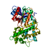
| Structure viewer | Molecule: MolmilJmol/JSmol |
|---|
- Downloads & links
-Download
| PDBx/mmCIF format | 3kdh.cif.gz | 257.5 KB | Display | PDBx/mmCIF format |
|---|---|---|---|---|
| PDB format | pdb3kdh.ent.gz | 208.8 KB | Display | PDB format |
| PDBx/mmJSON format | 3kdh.json.gz | Tree view | PDBx/mmJSON format | |
| Others |  Other downloads Other downloads |
-Validation report
| Arichive directory | https://data.pdbj.org/pub/pdb/validation_reports/kd/3kdhftp://data.pdbj.org/pub/pdb/validation_reports/kd/3kdh | HTTPS FTP |
|---|
-Related structure data


-Links
 PDBj
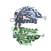
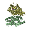
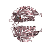
PDBj- Assembly
| Deposited unit | 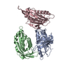
| |||||||||||||||||||||
|---|---|---|---|---|---|---|---|---|---|---|---|---|---|---|---|---|---|---|---|---|---|---|
| 1 | 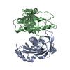
| |||||||||||||||||||||
| 2 | 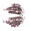
| |||||||||||||||||||||
| Unit cell |
| |||||||||||||||||||||
| Components on special symmetry positions |
|
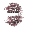
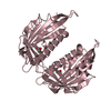
-Components
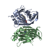
| #1: Protein | Mass: 21309.938 Da / Num. of mol.: 3 Source method: isolated from a genetically manipulated source Source: (gene. exp.)  #2: Water | ChemComp-HOH / |  Mass: 18.015 Da / Num. of mol.: 1022 / Source method: isolated from a natural source / Formula: H2O Mass: 18.015 Da / Num. of mol.: 1022 / Source method: isolated from a natural source / Formula: H2O |
|---|
-Experimental details
-Experiment
| Experiment | Method: X-RAY DIFFRACTION / Number of used crystals: 1 |
|---|
- Sample preparation
| Crystal | Density Matthews: 2.42 Å3/Da / Density % sol: 49.21 % |
|---|---|
| Crystal grow | Temperature: 291 K / Method: vapor diffusion, hanging drop / pH: 8.5 Details: 1M sodium citrate tribasic, 100mM Tris, pH8.5, 79mM MEGA-8 (Octanoyl-N-methylglucamide), VAPOR DIFFUSION, HANGING DROP, temperature 291K |
-Data collection
| Diffraction | Mean temperature: 100 K |
|---|---|
| Diffraction source | Source: SYNCHROTRON / Site: SPring-8  / Beamline: BL41XU / Wavelength: 1 Å / Beamline: BL41XU / Wavelength: 1 Å |
| Detector | Type: MARMOSAIC 225 mm CCD / Detector: CCD / Date: Jul 14, 2009 |
| Radiation | Protocol: SINGLE WAVELENGTH / Monochromatic (M) / Laue (L): M / Scattering type: x-ray |
| Radiation wavelength | Wavelength: 1 Å / Relative weight: 1 |
| Reflection | Resolution: 1.653→30.82 Å / Num. obs: 73929 / % possible obs: 98.6 % / Redundancy: 4 % / Biso Wilson estimate: 17.79 Å2 / Rmerge(I) obs: 0.041 / Net I/σ(I): 32.76 |
| Reflection shell | Resolution: 1.653→1.71 Å / Rmerge(I) obs: 0.211 / Mean I/σ(I) obs: 6.54 / % possible all: 94.4 |
- Processing
| Software |
| ||||||||||||||||||||||||||||||||||||||||||||||||||||||||||||||||||||||||||||||||||||||||||||||||||||||||||||||||||||||||||||||||||||||||||||||||||||||||||||||||||||||||||||||||||||||
|---|---|---|---|---|---|---|---|---|---|---|---|---|---|---|---|---|---|---|---|---|---|---|---|---|---|---|---|---|---|---|---|---|---|---|---|---|---|---|---|---|---|---|---|---|---|---|---|---|---|---|---|---|---|---|---|---|---|---|---|---|---|---|---|---|---|---|---|---|---|---|---|---|---|---|---|---|---|---|---|---|---|---|---|---|---|---|---|---|---|---|---|---|---|---|---|---|---|---|---|---|---|---|---|---|---|---|---|---|---|---|---|---|---|---|---|---|---|---|---|---|---|---|---|---|---|---|---|---|---|---|---|---|---|---|---|---|---|---|---|---|---|---|---|---|---|---|---|---|---|---|---|---|---|---|---|---|---|---|---|---|---|---|---|---|---|---|---|---|---|---|---|---|---|---|---|---|---|---|---|---|---|---|---|
| Refinement | Method to determine structure: SAD / Resolution: 1.653→30.82 Å / Occupancy max: 1 / Occupancy min: 0.16 / FOM work R set: 0.876 / SU ML: 0.2 / Isotropic thermal model: TLS / σ(F): 0.05 / Phase error: 19.57 / Stereochemistry target values: ML
| ||||||||||||||||||||||||||||||||||||||||||||||||||||||||||||||||||||||||||||||||||||||||||||||||||||||||||||||||||||||||||||||||||||||||||||||||||||||||||||||||||||||||||||||||||||||
| Solvent computation | Shrinkage radii: 0.9 Å / VDW probe radii: 1.11 Å / Solvent model: FLAT BULK SOLVENT MODEL / Bsol: 44.664 Å2 / ksol: 0.319 e/Å3 | ||||||||||||||||||||||||||||||||||||||||||||||||||||||||||||||||||||||||||||||||||||||||||||||||||||||||||||||||||||||||||||||||||||||||||||||||||||||||||||||||||||||||||||||||||||||
| Displacement parameters | Biso max: 107.6 Å2 / Biso mean: 26.027 Å2 / Biso min: 5.59 Å2
| ||||||||||||||||||||||||||||||||||||||||||||||||||||||||||||||||||||||||||||||||||||||||||||||||||||||||||||||||||||||||||||||||||||||||||||||||||||||||||||||||||||||||||||||||||||||
| Refinement step | Cycle: LAST / Resolution: 1.653→30.82 Å
| ||||||||||||||||||||||||||||||||||||||||||||||||||||||||||||||||||||||||||||||||||||||||||||||||||||||||||||||||||||||||||||||||||||||||||||||||||||||||||||||||||||||||||||||||||||||
| Refine LS restraints |
| ||||||||||||||||||||||||||||||||||||||||||||||||||||||||||||||||||||||||||||||||||||||||||||||||||||||||||||||||||||||||||||||||||||||||||||||||||||||||||||||||||||||||||||||||||||||
| LS refinement shell |
| ||||||||||||||||||||||||||||||||||||||||||||||||||||||||||||||||||||||||||||||||||||||||||||||||||||||||||||||||||||||||||||||||||||||||||||||||||||||||||||||||||||||||||||||||||||||
| Refinement TLS params. | Method: refined / Refine-ID: X-RAY DIFFRACTION
| ||||||||||||||||||||||||||||||||||||||||||||||||||||||||||||||||||||||||||||||||||||||||||||||||||||||||||||||||||||||||||||||||||||||||||||||||||||||||||||||||||||||||||||||||||||||
| Refinement TLS group |
|
