 Movie
Movie Controller

[English] 日本語
 Yorodumi
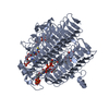
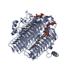
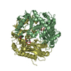
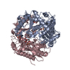
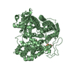
- PDB-3fsb: Crystal structure of QdtC, the dTDP-3-amino-3,6-dideoxy-D-glucose... -
+ Open data

- Basic information
| Entry | Database: PDB / ID: 3fsb | ||||||
|---|---|---|---|---|---|---|---|
| Title | Crystal structure of QdtC, the dTDP-3-amino-3,6-dideoxy-D-glucose N-acetyl transferase from Thermoanaerobacterium thermosaccharolyticum in complex with CoA and dTDP-3-amino-quinovose | ||||||
 Components Components | QdtC | ||||||
 Keywords Keywords | TRANSFERASE / N-acetyltransferase / deoxysugar / natural product | ||||||
| Function / homology |  Function and homology information Function and homology informationUbiquitin Ligase Nedd4; Chain: W; - #30 / Ubiquitin Ligase Nedd4; Chain: W; / Hexapeptide repeat of succinyl-transferase / : / Hexapeptide repeat proteins / UDP N-Acetylglucosamine Acyltransferase; domain 1 / Hexapeptide repeat / Other non-globular / Bacterial transferase hexapeptide (six repeats) / Trimeric LpxA-like superfamily ...Ubiquitin Ligase Nedd4; Chain: W; - #30 / Ubiquitin Ligase Nedd4; Chain: W; / Hexapeptide repeat of succinyl-transferase / : / Hexapeptide repeat proteins / UDP N-Acetylglucosamine Acyltransferase; domain 1 / Hexapeptide repeat / Other non-globular / Bacterial transferase hexapeptide (six repeats) / Trimeric LpxA-like superfamily / 3 Solenoid / Special / Mainly Beta Similarity search - Domain/homology | ||||||
| Biological species |  Thermoanaerobacterium thermosaccharolyticum (bacteria) Thermoanaerobacterium thermosaccharolyticum (bacteria) | ||||||
| Method |  X-RAY DIFFRACTION / MOLECULAR REPLACEMENT / Resolution: 1.95 Å X-RAY DIFFRACTION / MOLECULAR REPLACEMENT / Resolution: 1.95 Å | ||||||
 Authors Authors | Holden, H.M. / Thoden, J.B. | ||||||
 Citation Citation | Journal: Biochemistry / Year: 2009 Title: Structural and Functional Studies of QdtC: an N-Acetyltransferase Required for the Biosynthesis of dTDP-3-Acetamido-3,6-Dideoxy-alpha-D-Glucose. Authors: Thoden, J. / Cook, P. / Schaffer, C. / Messner, P. / Holden, H. | ||||||
| History |
|
- Structure visualization
| Structure viewer | Molecule: MolmilJmol/JSmol |
|---|
- Downloads & links
-Download
| PDBx/mmCIF format | 3fsb.cif.gz | 126.1 KB | Display | PDBx/mmCIF format |
|---|---|---|---|---|
| PDB format | pdb3fsb.ent.gz | 96.8 KB | Display | PDB format |
| PDBx/mmJSON format | 3fsb.json.gz | Tree view | PDBx/mmJSON format | |
| Others |  Other downloads Other downloads |
-Validation report
| Arichive directory | https://data.pdbj.org/pub/pdb/validation_reports/fs/3fsbftp://data.pdbj.org/pub/pdb/validation_reports/fs/3fsb | HTTPS FTP |
|---|
-Related structure data
| Related structure data |  3fs8SC  3fscC S: Starting model for refinement C: citing same article ( |
|---|---|
| Similar structure data |

-Links
 PDBj
PDBj


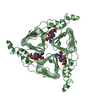
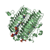
- Assembly
| Deposited unit | 
| |||||||||||||||||||||||||||
|---|---|---|---|---|---|---|---|---|---|---|---|---|---|---|---|---|---|---|---|---|---|---|---|---|---|---|---|---|
| 1 | 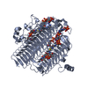
| |||||||||||||||||||||||||||
| 2 | 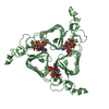
| |||||||||||||||||||||||||||
| Unit cell |
| |||||||||||||||||||||||||||
| Components on special symmetry positions |
|
-Components
| #1: Protein | Mass: 30816.162 Da / Num. of mol.: 2 Source method: isolated from a genetically manipulated source Source: (gene. exp.) Thermoanaerobacterium thermosaccharolyticum (bacteria)Strain: E2707-71 / Gene: qdtC / Plasmid: pET31 / Production host: #2: Chemical |   Mass: 767.534 Da / Num. of mol.: 2 / Source method: obtained synthetically / Formula: C21H36N7O16P3S Mass: 767.534 Da / Num. of mol.: 2 / Source method: obtained synthetically / Formula: C21H36N7O16P3S#3: Chemical | 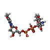  Mass: 547.345 Da / Num. of mol.: 2 / Source method: obtained synthetically / Formula: C16H27N3O14P2 Mass: 547.345 Da / Num. of mol.: 2 / Source method: obtained synthetically / Formula: C16H27N3O14P2#4: Water | ChemComp-HOH / |  Mass: 18.015 Da / Num. of mol.: 229 / Source method: isolated from a natural source / Formula: H2O Mass: 18.015 Da / Num. of mol.: 229 / Source method: isolated from a natural source / Formula: H2O |
|---|
-Experimental details
-Experiment
| Experiment | Method: X-RAY DIFFRACTION / Number of used crystals: 1 |
|---|
- Sample preparation
| Crystal | Density Matthews: 2.41 Å3/Da / Density % sol: 48.92 % |
|---|---|
| Crystal grow | Temperature: 298 K / Method: vapor diffusion, hanging drop / pH: 7 Details: 24% PEG 5000, 0.1M HEPES, 0.01 M Acetyl-CoA, 0.04M dTDP-3-aminoquinovose, 2% ethyleneglycol, pH 7.0, VAPOR DIFFUSION, HANGING DROP, temperature 298K |
-Data collection
| Diffraction | Mean temperature: 100 K |
|---|---|
| Diffraction source | Source: ROTATING ANODE / Type: RIGAKU RU200 / Wavelength: 1.5418 Å |
| Detector | Type: Bruker AXS Platinum135 / Detector: CCD / Date: Aug 1, 2008 / Details: Montel |
| Radiation | Monochromator: Ni filter / Protocol: SINGLE WAVELENGTH / Monochromatic (M) / Laue (L): M / Scattering type: x-ray |
| Radiation wavelength | Wavelength: 1.5418 Å / Relative weight: 1 |
| Reflection | Resolution: 1.95→30 Å / Num. all: 39913 / Num. obs: 39913 / % possible obs: 95.1 % / Observed criterion σ(F): 0 / Observed criterion σ(I): 0 / Redundancy: 3.4 % / Rsym value: 0.084 |
| Reflection shell | Resolution: 1.95→2.05 Å / Redundancy: 1.4 % / Mean I/σ(I) obs: 2.3 / Num. unique all: 4815 / Rsym value: 0.264 / % possible all: 79.9 |
- Processing
| Software |
| |||||||||||||||||||||||||
|---|---|---|---|---|---|---|---|---|---|---|---|---|---|---|---|---|---|---|---|---|---|---|---|---|---|---|
| Refinement | Method to determine structure: MOLECULAR REPLACEMENT Starting model: 3fs8 Resolution: 1.95→30 Å / σ(F): 0 / Stereochemistry target values: Engh & Huber
| |||||||||||||||||||||||||
| Refinement step | Cycle: LAST / Resolution: 1.95→30 Å
|
