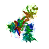
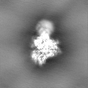
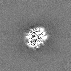
ジャーナル: Nat Commun / 年: 2023 タイトル: Structure of the DDB1-AMBRA1 E3 ligase receptor complex linked to cell cycle regulation. 著者: Ming Liu / Yang Wang / Fei Teng / Xinyi Mai / Xi Wang / Ming-Yuan Su / Goran Stjepanovic / 要旨: AMBRA1 is a tumor suppressor protein that functions as a substrate receptor of the ubiquitin conjugation system with roles in autophagy and the cell cycle regulatory network. The intrinsic disorder ...AMBRA1 is a tumor suppressor protein that functions as a substrate receptor of the ubiquitin conjugation system with roles in autophagy and the cell cycle regulatory network. The intrinsic disorder of AMBRA1 has thus far precluded its structural determination. To solve this problem, we analyzed the dynamics of AMBRA1 using hydrogen deuterium exchange mass spectrometry (HDX-MS). The HDX results indicated that AMBRA1 is a highly flexible protein and can be stabilized upon interaction with DDB1, the adaptor of the Cullin4A/B E3 ligase. Here, we present the cryo-EM structure of AMBRA1 in complex with DDB1 at 3.08 Å resolution. The structure shows that parts of the N- and C-terminal structural regions in AMBRA1 fold together into the highly dynamic WD40 domain and reveals how DDB1 engages with AMBRA1 to create a binding scaffold for substrate recruitment. The N-terminal helix-loop-helix motif and WD40 domain of AMBRA1 associate with the double-propeller fold of DDB1. We also demonstrate that DDB1 binding-defective AMBRA1 mutants prevent ubiquitination of the substrate Cyclin D1 in vitro and increase cell cycle progression. Together, these results provide structural insights into the AMBRA1-ubiquitin ligase complex and suggest a mechanism by which AMBRA1 acts as a hub involved in various physiological processes.
 ムービー
ムービー コントローラー


 データを開く
 基本情報
基本情報
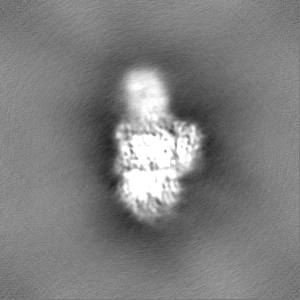
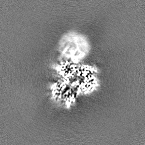
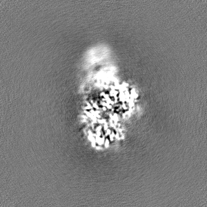
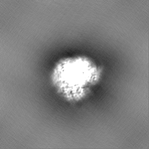
 マップデータ
 試料
試料 キーワード
キーワード E3 ligase (ユビキチンリガーゼ) /
  機能・相同性情報
 データ登録者
 中国, 1件
  引用
引用 構造の表示
構造の表示
 ダウンロードとリンク
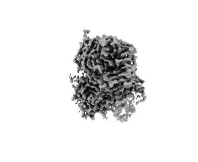 emd_37752.png
 http://ftp.pdbj.org/pub/emdb/structures/EMD-37752


 Z
Z Y
Y X
X


 試料の構成要素
 解析
解析 電子顕微鏡法