 Movie
Movie Controller

[English] 日本語
 Yorodumi
- PDB-8wqr: Structure of the DDB1-AMBRA1 E3 ligase receptor complex linked to... -
+ Open data

- Basic information
| Entry | Database: PDB / ID: 8wqr | |||||||||||||||||||||
|---|---|---|---|---|---|---|---|---|---|---|---|---|---|---|---|---|---|---|---|---|---|---|
| Title | Structure of the DDB1-AMBRA1 E3 ligase receptor complex linked to cell cycle regulation | |||||||||||||||||||||
 Components Components |
| |||||||||||||||||||||
 Keywords Keywords | STRUCTURAL PROTEIN / E3 ligase | |||||||||||||||||||||
| Function / homology |  Function and homology information Function and homology informationpositive regulation of free ubiquitin chain polymerization / response to mitochondrial depolarisation / positive regulation of mitophagy / positive regulation by virus of viral protein levels in host cell / spindle assembly involved in female meiosis / neural tube development / epigenetic programming in the zygotic pronuclei / positive regulation of regulatory T cell differentiation / UV-damage excision repair / biological process involved in interaction with symbiont ...positive regulation of free ubiquitin chain polymerization / response to mitochondrial depolarisation / positive regulation of mitophagy / positive regulation by virus of viral protein levels in host cell / spindle assembly involved in female meiosis / neural tube development / epigenetic programming in the zygotic pronuclei / positive regulation of regulatory T cell differentiation / UV-damage excision repair / biological process involved in interaction with symbiont / regulation of mitotic cell cycle phase transition / Macroautophagy / WD40-repeat domain binding / Cul4A-RING E3 ubiquitin ligase complex / Cul4-RING E3 ubiquitin ligase complex / Cul4B-RING E3 ubiquitin ligase complex / ubiquitin ligase complex scaffold activity / negative regulation of reproductive process / negative regulation of developmental process / negative regulation of cardiac muscle cell apoptotic process / regulation of G1/S transition of mitotic cell cycle / viral release from host cell / protein phosphatase activator activity / cullin family protein binding / axoneme / ectopic germ cell programmed cell death / positive regulation of viral genome replication / autophagosome assembly / mitophagy / ubiquitin-like ligase-substrate adaptor activity / phagocytic vesicle / proteasomal protein catabolic process / positive regulation of autophagy / autophagosome / positive regulation of gluconeogenesis / cellular response to starvation / nucleotide-excision repair / sperm end piece / regulation of circadian rhythm / Recognition of DNA damage by PCNA-containing replication complex / DNA Damage Recognition in GG-NER / Dual Incision in GG-NER / Transcription-Coupled Nucleotide Excision Repair (TC-NER) / Wnt signaling pathway / Formation of TC-NER Pre-Incision Complex / Formation of Incision Complex in GG-NER / protein polyubiquitination / Dual incision in TC-NER / positive regulation of protein catabolic process / Gap-filling DNA repair synthesis and ligation in TC-NER / cellular response to UV / rhythmic process / site of double-strand break / sperm principal piece / Neddylation / GTPase binding / sperm midpiece / protein phosphatase binding / ubiquitin-dependent protein catabolic process / cytoskeleton / damaged DNA binding / negative regulation of neuron apoptotic process / proteasome-mediated ubiquitin-dependent protein catabolic process / protein-macromolecule adaptor activity / cell differentiation / mitochondrial outer membrane / chromosome, telomeric region / positive regulation of phosphatidylinositol 3-kinase/protein kinase B signal transduction / protein ubiquitination / negative regulation of cell population proliferation / focal adhesion / DNA repair / apoptotic process / DNA damage response / ubiquitin protein ligase binding / regulation of transcription by RNA polymerase II / negative regulation of apoptotic process / protein-containing complex binding / nucleolus / perinuclear region of cytoplasm / endoplasmic reticulum / protein-containing complex / mitochondrion / : / DNA binding / extracellular exosome / nucleoplasm / nucleus / cytoplasm / cytosol Similarity search - Function | |||||||||||||||||||||
| Biological species |  Homo sapiens (human) Homo sapiens (human) | |||||||||||||||||||||
| Method | ELECTRON MICROSCOPY / single particle reconstruction / cryo EM / Resolution: 3.08 Å | |||||||||||||||||||||
 Authors Authors | Liu, M. / Wang, Y. / Su, M.Y. / Stjepanovic, G. | |||||||||||||||||||||
| Funding support |  China, 1items China, 1items
| |||||||||||||||||||||
 Citation Citation | Journal: Nat Commun / Year: 2023 Title: Structure of the DDB1-AMBRA1 E3 ligase receptor complex linked to cell cycle regulation. Authors: Ming Liu / Yang Wang / Fei Teng / Xinyi Mai / Xi Wang / Ming-Yuan Su / Goran Stjepanovic / Abstract: AMBRA1 is a tumor suppressor protein that functions as a substrate receptor of the ubiquitin conjugation system with roles in autophagy and the cell cycle regulatory network. The intrinsic disorder ...AMBRA1 is a tumor suppressor protein that functions as a substrate receptor of the ubiquitin conjugation system with roles in autophagy and the cell cycle regulatory network. The intrinsic disorder of AMBRA1 has thus far precluded its structural determination. To solve this problem, we analyzed the dynamics of AMBRA1 using hydrogen deuterium exchange mass spectrometry (HDX-MS). The HDX results indicated that AMBRA1 is a highly flexible protein and can be stabilized upon interaction with DDB1, the adaptor of the Cullin4A/B E3 ligase. Here, we present the cryo-EM structure of AMBRA1 in complex with DDB1 at 3.08 Å resolution. The structure shows that parts of the N- and C-terminal structural regions in AMBRA1 fold together into the highly dynamic WD40 domain and reveals how DDB1 engages with AMBRA1 to create a binding scaffold for substrate recruitment. The N-terminal helix-loop-helix motif and WD40 domain of AMBRA1 associate with the double-propeller fold of DDB1. We also demonstrate that DDB1 binding-defective AMBRA1 mutants prevent ubiquitination of the substrate Cyclin D1 in vitro and increase cell cycle progression. Together, these results provide structural insights into the AMBRA1-ubiquitin ligase complex and suggest a mechanism by which AMBRA1 acts as a hub involved in various physiological processes. | |||||||||||||||||||||
| History |
|
- Structure visualization
| Structure viewer | Molecule: MolmilJmol/JSmol |
|---|
- Downloads & links
-Download
| PDBx/mmCIF format | 8wqr.cif.gz | 242.7 KB | Display | PDBx/mmCIF format |
|---|---|---|---|---|
| PDB format | pdb8wqr.ent.gz | 181.8 KB | Display | PDB format |
| PDBx/mmJSON format | 8wqr.json.gz | Tree view | PDBx/mmJSON format | |
| Others |  Other downloads Other downloads |
-Validation report
| Arichive directory | https://data.pdbj.org/pub/pdb/validation_reports/wq/8wqrftp://data.pdbj.org/pub/pdb/validation_reports/wq/8wqr | HTTPS FTP |
|---|
-Related structure data
| Related structure data |  37752MC M: map data used to model this data C: citing same article ( |
|---|---|
| Similar structure data |
-Links
 PDBj
PDBj


- Assembly
| Deposited unit | 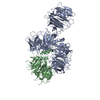
|
|---|---|
| 1 |
|
-Components
| #1: Protein | Mass: 44496.871 Da / Num. of mol.: 1 Source method: isolated from a genetically manipulated source Source: (gene. exp.) Homo sapiens (human) / Gene: AMBRA1 / Production host: Homo sapiens (human) / References: UniProt: Q9C0C7 |
|---|---|
| #2: Protein | Mass: 127097.469 Da / Num. of mol.: 1 Source method: isolated from a genetically manipulated source Source: (gene. exp.) Homo sapiens (human) / Gene: DDB1, XAP1 / Production host: Homo sapiens (human) / References: UniProt: Q16531 |
| Has protein modification | N |
-Experimental details
-Experiment
| Experiment | Method: ELECTRON MICROSCOPY |
|---|---|
| EM experiment | Aggregation state: PARTICLE / 3D reconstruction method: single particle reconstruction |
- Sample preparation
| Component | Name: DDB1-AMBRA1 complex / Type: COMPLEX / Entity ID: all / Source: RECOMBINANT |
|---|---|
| Molecular weight | Experimental value: NO |
| Source (natural) | Organism: Homo sapiens (human) |
| Source (recombinant) | Organism: Homo sapiens (human) |
| Buffer solution | pH: 7.4 |
| Specimen | Embedding applied: NO / Shadowing applied: NO / Staining applied: NO / Vitrification applied: YES |
| Vitrification | Cryogen name: ETHANE |
- Electron microscopy imaging
| Experimental equipment |  Model: Titan Krios / Image courtesy: FEI Company |
|---|---|
| Microscopy | Model: FEI TITAN KRIOS Details: Grid information as following: Company/model: Quantifoil Cu 1.2/1.3 Material:Cu Grid mesh: 200 |
| Electron gun | Electron source:  FIELD EMISSION GUN / Accelerating voltage: 300 kV / Illumination mode: SPOT SCAN FIELD EMISSION GUN / Accelerating voltage: 300 kV / Illumination mode: SPOT SCAN |
| Electron lens | Mode: BRIGHT FIELD / Nominal defocus max: 1800 nm / Nominal defocus min: 1000 nm |
| Image recording | Electron dose: 58.96 e/Å2 / Film or detector model: GATAN K3 (6k x 4k) |
- Processing
| EM software | Name: PHENIX / Category: model refinement | ||||||||||||||||||||||||
|---|---|---|---|---|---|---|---|---|---|---|---|---|---|---|---|---|---|---|---|---|---|---|---|---|---|
| CTF correction | Type: NONE | ||||||||||||||||||||||||
| 3D reconstruction | Resolution: 3.08 Å / Resolution method: FSC 0.143 CUT-OFF / Num. of particles: 780090 / Symmetry type: POINT | ||||||||||||||||||||||||
| Refine LS restraints |
|
