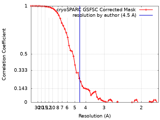
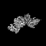
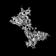
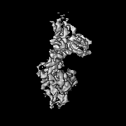
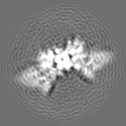
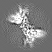
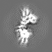
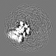
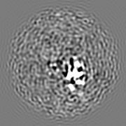
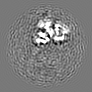
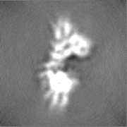
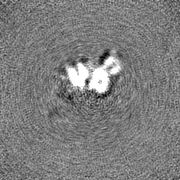
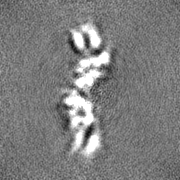
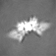
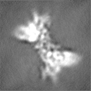
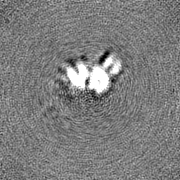
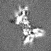
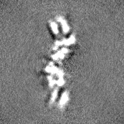
Journal: Exploration (Beijing) / Year: 2024 Title: A novel nanobody broadly neutralizes SARS-CoV-2 via induction of spike trimer dimers conformation. Authors: Yang Yang / Junfang Zhang / Shengnan Zhang / Chenhui Zhang / Chenguang Shen / Shuo Song / Yanqun Wang / Yun Peng / Xiaohua Gong / Jun Dai / Chongwei Xie / Tatyana Aleksandrovna Khrustaleva / ...Authors: Yang Yang / Junfang Zhang / Shengnan Zhang / Chenhui Zhang / Chenguang Shen / Shuo Song / Yanqun Wang / Yun Peng / Xiaohua Gong / Jun Dai / Chongwei Xie / Tatyana Aleksandrovna Khrustaleva / Vladislav Victorovich Khrustalev / Yongting Huo / Di Lu / Da Yao / Jincun Zhao / Yingxia Liu / Hongzhou Lu / Abstract: The ongoing mutations of the SARS-CoV-2 pose serious challenges to the efficacy of the available antiviral drugs, and new drugs with fantastic efficacy are always deserved investigation. Here, a ...The ongoing mutations of the SARS-CoV-2 pose serious challenges to the efficacy of the available antiviral drugs, and new drugs with fantastic efficacy are always deserved investigation. Here, a nanobody called IBT-CoV144 is reported, which exhibits broad neutralizing activity against SARS-CoV-2 by inducing the conformation of spike trimer dimers. IBT-CoV144 was isolated from an immunized alpaca using the RBD of wild-type SARS-CoV-2, and it showed strong cross-reactive binding and neutralizing potency against diverse SARS-CoV-2 variants, including Omicron subvariants. Moreover, the prophylactically and therapeutically intranasal administration of IBT-CoV144 confers fantastic protective efficacy against the challenge of Omicron BA.1 variant in BALB/c mice model. The structure analysis of the complex between spike (S) protein, conducted using Cryo-EM, revealed a special conformation known as the trimer dimers. This conformation is formed by two trimers, with six RBDs in the "up" state and bound by six VHHs. IBT-CoV144 binds to the lateral region of the RBD on the S protein, facilitating the aggregation of S proteins. This aggregation results in steric hindrance, which disrupts the recognition of the virus by ACE2 on host cells. The discovery of IBT-CoV144 will provide valuable insights for the development of advanced therapeutics and the design of next-generation vaccines.
In the structure databanks used in Yorodumi, some data are registered as the other names, "COVID-19 virus" and "2019-nCoV". Here are the details of the virus and the list of structure data.
Jan 31, 2019. EMDB accession codes are about to change! (news from PDBe EMDB page)
EMDB accession codes are about to change! (news from PDBe EMDB page)
The allocation of 4 digits for EMDB accession codes will soon come to an end. Whilst these codes will remain in use, new EMDB accession codes will include an additional digit and will expand incrementally as the available range of codes is exhausted. The current 4-digit format prefixed with “EMD-” (i.e. EMD-XXXX) will advance to a 5-digit format (i.e. EMD-XXXXX), and so on. It is currently estimated that the 4-digit codes will be depleted around Spring 2019, at which point the 5-digit format will come into force.
The EM Navigator/Yorodumi systems omit the EMD- prefix.
Related info.:Q: What is EMD? / ID/Accession-code notation in Yorodumi/EM Navigator
Yorodumi is a browser for structure data from EMDB, PDB, SASBDB, etc.
This page is also the successor to EM Navigator detail page, and also detail information page/front-end page for Omokage search.
The word "yorodu" (or yorozu) is an old Japanese word meaning "ten thousand". "mi" (miru) is to see.
Related info.:EMDB / PDB / SASBDB / Comparison of 3 databanks / Yorodumi Search / Aug 31, 2016. New EM Navigator & Yorodumi / Yorodumi Papers / Jmol/JSmol / Function and homology information / Changes in new EM Navigator and Yorodumi
 Movie
Movie Controller

 Yorodumi
 Open data
 Basic information
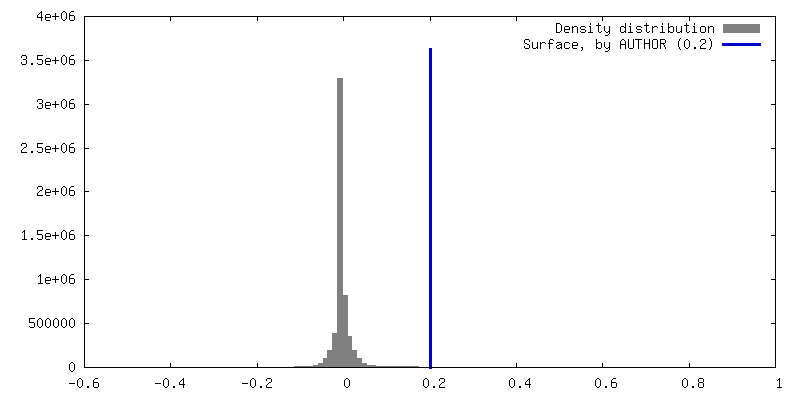
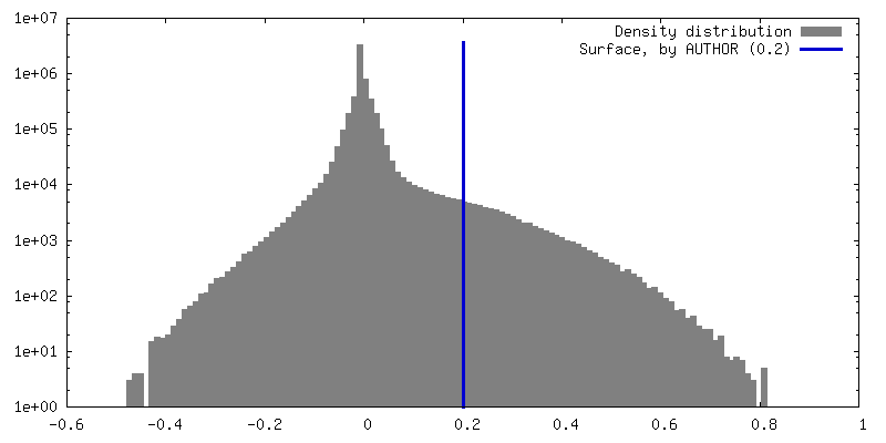
 Map data
 Sample
 Keywords
 Function and homology information

 Severe acute respiratory syndrome coronavirus 2
 Authors
 China, 1 items
  Citation
 Structure visualization
 Downloads & links
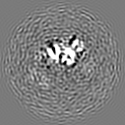
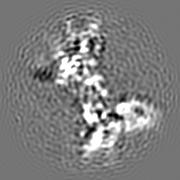
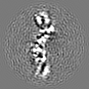
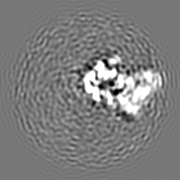
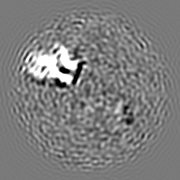
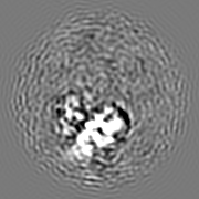
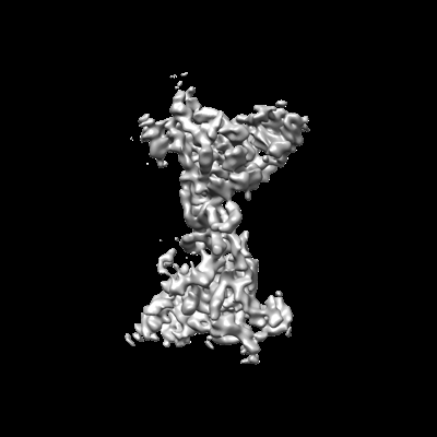 emd_36730.png
 http://ftp.pdbj.org/pub/emdb/structures/EMD-36730


 Z (Sec.)
 Y (Row.)
 X (Col.)


 Sample components
 Homo sapiens (human)
 Processing
 Electron microscopy
 FIELD EMISSION GUN