 ムービー
ムービー コントローラー


+ データを開く

- 基本情報
基本情報
| 登録情報 | データベース: EMDB / ID: EMD-3557 | |||||||||
|---|---|---|---|---|---|---|---|---|---|---|
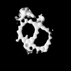
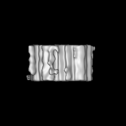
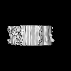
| タイトル | Centriolar Distal MT doublets in centrosome extracted from o.aries thymocytes | |||||||||
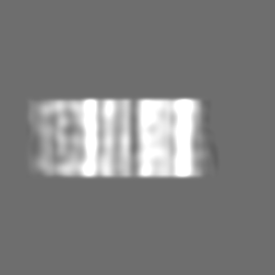
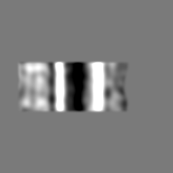
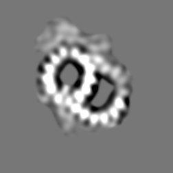
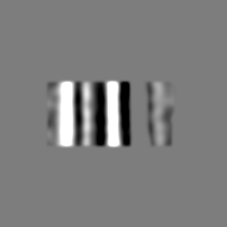
 マップデータ マップデータ | MT doublet at the distal part of the centriolar wall of centrosome in mammals. The subtomogram averaging was processed using tomograms of centrosomes extracted from o.aries thymocytes | |||||||||
 試料 試料 |
| |||||||||
| 生物種 |  | |||||||||
| 手法 | サブトモグラム平均法 / クライオ電子顕微鏡法 / 解像度: 46.7 Å | |||||||||
 データ登録者 データ登録者 | Busselez J / Chichon FJ / Melero R / Carrascosa JL / Carazo JM | |||||||||
 引用 引用 | ジャーナル: Sci Rep / 年: 2019 タイトル: Cryo-Electron Tomography and Proteomics studies of centrosomes from differentiated quiescent thymocytes. 著者: Johan Busselez / Francisco Javier Chichón / Maria Josefa Rodríguez / Adan Alpízar / Séverine Isabelle Gharbi / Mònica Franch / Roberto Melero / Alberto Paradela / José L Carrascosa / José-Maria Carazo /   要旨: We have used cryo Electron Tomography, proteomics and immunolabeling to study centrosomes isolated from the young lamb thymus, an efficient source of quiescent differentiated cells. We compared the ...We have used cryo Electron Tomography, proteomics and immunolabeling to study centrosomes isolated from the young lamb thymus, an efficient source of quiescent differentiated cells. We compared the proteome of thymocyte centrosomes to data published for KE37 cells, focusing on proteins associated with centriole disengagement and centrosome separation. The data obtained enhances our understanding of the protein system joining the centrioles, a system comprised of a branched network of fibers linked to an apparently amorphous density that was partially characterized here. A number of proteins were localized to the amorphous density by immunolabeling (C-NAP1, cohesin SMC1, condensin SMC4 and NCAPD2), yet not DNA. In conjuction, these data not only extend our understanding of centrosomes but they will help refine the model that focus on the protein system associated with the centriolar junction. | |||||||||
| 履歴 |
|
- 構造の表示
構造の表示
| ムービー |
 ムービービューア ムービービューア |
|---|---|
| 構造ビューア | EMマップ: SurfViewMolmilJmol/JSmol |
| 添付画像 |

- ダウンロードとリンク
-EMDBアーカイブ
| マップデータ | emd_3557.map.gz | 5.8 MB | EMDBマップデータ形式 | |
|---|---|---|---|---|
| ヘッダ (付随情報) | emd-3557-v30.xmlemd-3557.xml | 14.7 KB 14.7 KB | 表示 表示 | EMDBヘッダ |
| FSC (解像度算出) | emd_3557_fsc.xml | 9.1 KB | 表示 | FSCデータファイル |
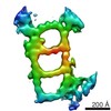
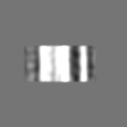
| 画像 | 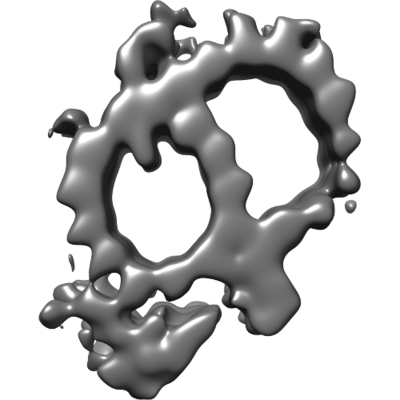 emd_3557.png emd_3557.png | 51.7 KB | ||
| アーカイブディレクトリ |  http://ftp.pdbj.org/pub/emdb/structures/EMD-3557ftp://ftp.pdbj.org/pub/emdb/structures/EMD-3557 http://ftp.pdbj.org/pub/emdb/structures/EMD-3557ftp://ftp.pdbj.org/pub/emdb/structures/EMD-3557 | HTTPS FTP |
-検証レポート
| 文書・要旨 | emd_3557_validation.pdf.gz | 229.1 KB | 表示 | EMDB検証レポート |
|---|---|---|---|---|
| 文書・詳細版 | emd_3557_full_validation.pdf.gz | 228.2 KB | 表示 | |
| XML形式データ | emd_3557_validation.xml.gz | 11.4 KB | 表示 | |
| アーカイブディレクトリ | https://ftp.pdbj.org/pub/emdb/validation_reports/EMD-3557ftp://ftp.pdbj.org/pub/emdb/validation_reports/EMD-3557 | HTTPS FTP |
-関連構造データ

-リンク
| EMDBのページ | EMDB (EBI/PDBe) / EMDataResource |
|---|
-マップ
| ファイル | ダウンロード / ファイル: emd_3557.map.gz / 形式: CCP4 / 大きさ: 61 MB / タイプ: IMAGE STORED AS FLOATING POINT NUMBER (4 BYTES) | ||||||||||||||||||||||||||||||||||||||||||||||||||||||||||||
|---|---|---|---|---|---|---|---|---|---|---|---|---|---|---|---|---|---|---|---|---|---|---|---|---|---|---|---|---|---|---|---|---|---|---|---|---|---|---|---|---|---|---|---|---|---|---|---|---|---|---|---|---|---|---|---|---|---|---|---|---|---|
| 注釈 | MT doublet at the distal part of the centriolar wall of centrosome in mammals. The subtomogram averaging was processed using tomograms of centrosomes extracted from o.aries thymocytes | ||||||||||||||||||||||||||||||||||||||||||||||||||||||||||||
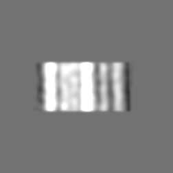
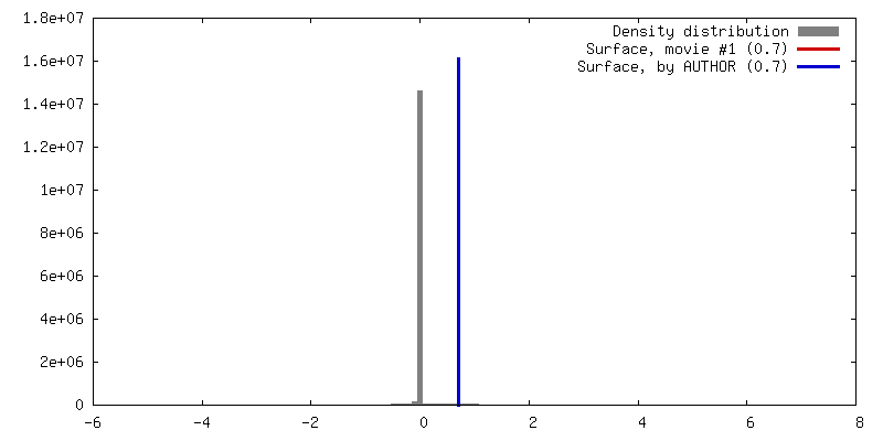
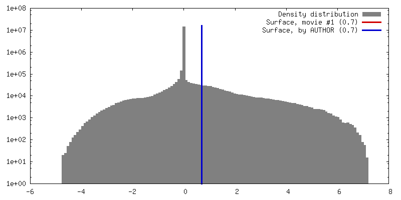
| 投影像・断面図 | 画像のコントロール
画像は Spider により作成 | ||||||||||||||||||||||||||||||||||||||||||||||||||||||||||||
| ボクセルのサイズ | X=Y=Z: 3.42 Å | ||||||||||||||||||||||||||||||||||||||||||||||||||||||||||||
| 密度 |
| ||||||||||||||||||||||||||||||||||||||||||||||||||||||||||||
| 対称性 | 空間群: 1 | ||||||||||||||||||||||||||||||||||||||||||||||||||||||||||||
| 詳細 | EMDB XML:
CCP4マップ ヘッダ情報:
| ||||||||||||||||||||||||||||||||||||||||||||||||||||||||||||
 Z (Sec.)
 Y (Row.)
 X (Col.)


-添付データ
- 試料の構成要素
-全体 : Centrosomes enriched from young lamb thymocyte
| 全体 | 名称: Centrosomes enriched from young lamb thymocyte |
|---|---|
| 要素 |
|
-超分子 #1: Centrosomes enriched from young lamb thymocyte
| 超分子 | 名称: Centrosomes enriched from young lamb thymocyte / タイプ: organelle_or_cellular_component / ID: 1 / 親要素: 0 詳細: The centrosomes were extracted accordingly to Komesli et. al. 1989 |
|---|---|
| 由来(天然) | 生物種: |
| 分子量 | 実験値: 230000 MDa |
-超分子 #2: Centriole
| 超分子 | 名称: Centriole / タイプ: organelle_or_cellular_component / ID: 2 / 親要素: 1 |
|---|---|
| 由来(天然) | 生物種: |
-実験情報
-構造解析
| 手法 | クライオ電子顕微鏡法 |
|---|---|
 解析 解析 | サブトモグラム平均法 |
| 試料の集合状態 | particle |
-試料調製
| 濃度 | 0.076 mg/mL |
|---|---|
| 緩衝液 | pH: 7.2 / 構成要素 - 濃度: 10.0 mM / 構成要素 - 式: C8H18N2O6S2 / 構成要素 - 名称: Pipes / 詳細: pH Rectified with KOH |
| グリッド | モデル: quantifoil 3.5/1 / 材質: COPPER/RHODIUM / メッシュ: 300 / 支持フィルム - 材質: FORMVAR / 支持フィルム - トポロジー: HOLEY |
| 凍結 | 凍結剤: ETHANE / 装置: LEICA EM CPC |
| 詳細 | The sample was enriched by two step of sedimentation in sucrose gradient. The sucrose was washed out in several drops of buffer, the last of which included fiducial markers (10 nm bovine serum albumin (BSA)-coated gold beads) for tilt series alignment |
- 電子顕微鏡法
| 顕微鏡 | FEI TITAN KRIOS |
|---|---|
| 特殊光学系 | エネルギーフィルター - 名称: Gatan 968 Quantum エネルギーフィルター - エネルギー下限: -10 eV エネルギーフィルター - エネルギー上限: 10 eV |
| 撮影 | フィルム・検出器のモデル: GATAN K2 QUANTUM (4k x 4k) デジタル化 - サイズ - 横: 3838 pixel / デジタル化 - サイズ - 縦: 3710 pixel / 平均露光時間: 1.0 sec. / 平均電子線量: 1.4 e/Å2 |
| 電子線 | 加速電圧: 300 kV / 電子線源:  FIELD EMISSION GUN FIELD EMISSION GUN |
| 電子光学系 | 最大 デフォーカス(補正後): 5.906 µm / 最小 デフォーカス(補正後): 5.54 µm / 照射モード: OTHER / 撮影モード: BRIGHT FIELD / Cs: 2.0 mm / 最大 デフォーカス(公称値): 5.0 µm / 最小 デフォーカス(公称値): 5.0 µm / 倍率(公称値): 42000 |
| 試料ステージ | 試料ホルダーモデル: FEI TITAN KRIOS AUTOGRID HOLDER ホルダー冷却材: NITROGEN |
| 実験機器 |  モデル: Titan Krios / 画像提供: FEI Company |

