 Movie
Movie Controller

[English] 日本語
 Yorodumi
- EMDB-3557: Centriolar Distal MT doublets in centrosome extracted from o.arie... -
+ Open data

- Basic information
| Entry | Database: EMDB / ID: EMD-3557 | |||||||||
|---|---|---|---|---|---|---|---|---|---|---|
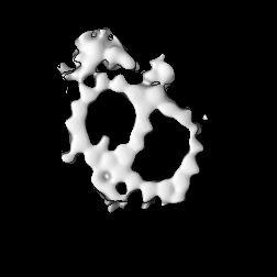
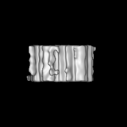
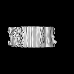
| Title | Centriolar Distal MT doublets in centrosome extracted from o.aries thymocytes | |||||||||
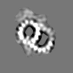
 Map data Map data | MT doublet at the distal part of the centriolar wall of centrosome in mammals. The subtomogram averaging was processed using tomograms of centrosomes extracted from o.aries thymocytes | |||||||||
 Sample Sample |
| |||||||||
| Biological species |  | |||||||||
| Method | subtomogram averaging / cryo EM / Resolution: 46.7 Å | |||||||||
 Authors Authors | Busselez J / Chichon FJ / Melero R / Carrascosa JL / Carazo JM | |||||||||
 Citation Citation | Journal: Sci Rep / Year: 2019 Title: Cryo-Electron Tomography and Proteomics studies of centrosomes from differentiated quiescent thymocytes. Authors: Johan Busselez / Francisco Javier Chichón / Maria Josefa Rodríguez / Adan Alpízar / Séverine Isabelle Gharbi / Mònica Franch / Roberto Melero / Alberto Paradela / José L Carrascosa / José-Maria Carazo /   Abstract: We have used cryo Electron Tomography, proteomics and immunolabeling to study centrosomes isolated from the young lamb thymus, an efficient source of quiescent differentiated cells. We compared the ...We have used cryo Electron Tomography, proteomics and immunolabeling to study centrosomes isolated from the young lamb thymus, an efficient source of quiescent differentiated cells. We compared the proteome of thymocyte centrosomes to data published for KE37 cells, focusing on proteins associated with centriole disengagement and centrosome separation. The data obtained enhances our understanding of the protein system joining the centrioles, a system comprised of a branched network of fibers linked to an apparently amorphous density that was partially characterized here. A number of proteins were localized to the amorphous density by immunolabeling (C-NAP1, cohesin SMC1, condensin SMC4 and NCAPD2), yet not DNA. In conjuction, these data not only extend our understanding of centrosomes but they will help refine the model that focus on the protein system associated with the centriolar junction. | |||||||||
| History |
|
- Structure visualization
| Movie |
 Movie viewer Movie viewer |
|---|---|
| Structure viewer | EM map: SurfViewMolmilJmol/JSmol |
| Supplemental images |

- Downloads & links
-EMDB archive
| Map data | emd_3557.map.gz | 5.8 MB | EMDB map data format | |
|---|---|---|---|---|
| Header (meta data) | emd-3557-v30.xmlemd-3557.xml | 14.7 KB 14.7 KB | Display Display | EMDB header |
| FSC (resolution estimation) | emd_3557_fsc.xml | 9.1 KB | Display | FSC data file |
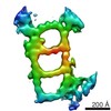
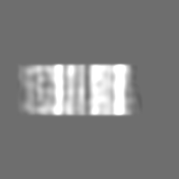
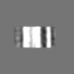
| Images | 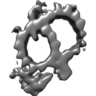 emd_3557.png emd_3557.png | 51.7 KB | ||
| Archive directory |  http://ftp.pdbj.org/pub/emdb/structures/EMD-3557ftp://ftp.pdbj.org/pub/emdb/structures/EMD-3557 http://ftp.pdbj.org/pub/emdb/structures/EMD-3557ftp://ftp.pdbj.org/pub/emdb/structures/EMD-3557 | HTTPS FTP |
-Validation report
| Summary document | emd_3557_validation.pdf.gz | 229.1 KB | Display | EMDB validaton report |
|---|---|---|---|---|
| Full document | emd_3557_full_validation.pdf.gz | 228.2 KB | Display | |
| Data in XML | emd_3557_validation.xml.gz | 11.4 KB | Display | |
| Arichive directory | https://ftp.pdbj.org/pub/emdb/validation_reports/EMD-3557ftp://ftp.pdbj.org/pub/emdb/validation_reports/EMD-3557 | HTTPS FTP |
-Related structure data

-Links
| EMDB pages | EMDB (EBI/PDBe) / EMDataResource |
|---|
-Map
| File | Download / File: emd_3557.map.gz / Format: CCP4 / Size: 61 MB / Type: IMAGE STORED AS FLOATING POINT NUMBER (4 BYTES) | ||||||||||||||||||||||||||||||||||||||||||||||||||||||||||||
|---|---|---|---|---|---|---|---|---|---|---|---|---|---|---|---|---|---|---|---|---|---|---|---|---|---|---|---|---|---|---|---|---|---|---|---|---|---|---|---|---|---|---|---|---|---|---|---|---|---|---|---|---|---|---|---|---|---|---|---|---|---|
| Annotation | MT doublet at the distal part of the centriolar wall of centrosome in mammals. The subtomogram averaging was processed using tomograms of centrosomes extracted from o.aries thymocytes | ||||||||||||||||||||||||||||||||||||||||||||||||||||||||||||
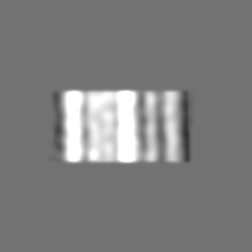
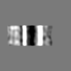
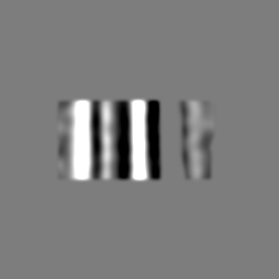
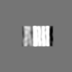
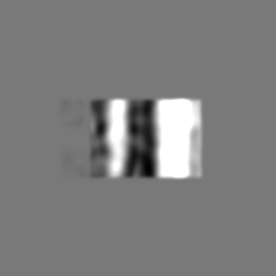
| Projections & slices | Image control
Images are generated by Spider. | ||||||||||||||||||||||||||||||||||||||||||||||||||||||||||||
| Voxel size | X=Y=Z: 3.42 Å | ||||||||||||||||||||||||||||||||||||||||||||||||||||||||||||
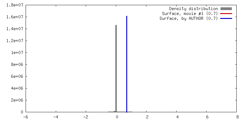
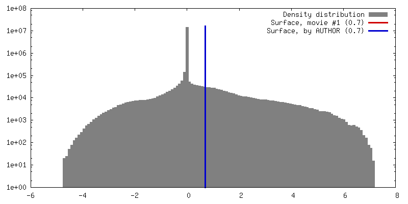
| Density |
| ||||||||||||||||||||||||||||||||||||||||||||||||||||||||||||
| Symmetry | Space group: 1 | ||||||||||||||||||||||||||||||||||||||||||||||||||||||||||||
| Details | EMDB XML:
CCP4 map header:
| ||||||||||||||||||||||||||||||||||||||||||||||||||||||||||||
 Z (Sec.)
 Y (Row.)
 X (Col.)


-Supplemental data
- Sample components
-Entire : Centrosomes enriched from young lamb thymocyte
| Entire | Name: Centrosomes enriched from young lamb thymocyte |
|---|---|
| Components |
|
-Supramolecule #1: Centrosomes enriched from young lamb thymocyte
| Supramolecule | Name: Centrosomes enriched from young lamb thymocyte / type: organelle_or_cellular_component / ID: 1 / Parent: 0 Details: The centrosomes were extracted accordingly to Komesli et. al. 1989 |
|---|---|
| Source (natural) | Organism: |
| Molecular weight | Experimental: 230000 MDa |
-Supramolecule #2: Centriole
| Supramolecule | Name: Centriole / type: organelle_or_cellular_component / ID: 2 / Parent: 1 |
|---|---|
| Source (natural) | Organism: |
-Experimental details
-Structure determination
| Method | cryo EM |
|---|---|
 Processing Processing | subtomogram averaging |
| Aggregation state | particle |
-Sample preparation
| Concentration | 0.076 mg/mL |
|---|---|
| Buffer | pH: 7.2 / Component - Concentration: 10.0 mM / Component - Formula: C8H18N2O6S2 / Component - Name: Pipes / Details: pH Rectified with KOH |
| Grid | Model: quantifoil 3.5/1 / Material: COPPER/RHODIUM / Mesh: 300 / Support film - Material: FORMVAR / Support film - topology: HOLEY |
| Vitrification | Cryogen name: ETHANE / Instrument: LEICA EM CPC |
| Details | The sample was enriched by two step of sedimentation in sucrose gradient. The sucrose was washed out in several drops of buffer, the last of which included fiducial markers (10 nm bovine serum albumin (BSA)-coated gold beads) for tilt series alignment |
- Electron microscopy
| Microscope | FEI TITAN KRIOS |
|---|---|
| Specialist optics | Energy filter - Name: Gatan 968 Quantum / Energy filter - Lower energy threshold: -10 eV / Energy filter - Upper energy threshold: 10 eV |
| Image recording | Film or detector model: GATAN K2 QUANTUM (4k x 4k) / Digitization - Dimensions - Width: 3838 pixel / Digitization - Dimensions - Height: 3710 pixel / Average exposure time: 1.0 sec. / Average electron dose: 1.4 e/Å2 |
| Electron beam | Acceleration voltage: 300 kV / Electron source:  FIELD EMISSION GUN FIELD EMISSION GUN |
| Electron optics | Calibrated defocus max: 5.906 µm / Calibrated defocus min: 5.54 µm / Illumination mode: OTHER / Imaging mode: BRIGHT FIELD / Cs: 2.0 mm / Nominal defocus max: 5.0 µm / Nominal defocus min: 5.0 µm / Nominal magnification: 42000 |
| Sample stage | Specimen holder model: FEI TITAN KRIOS AUTOGRID HOLDER / Cooling holder cryogen: NITROGEN |
| Experimental equipment |  Model: Titan Krios / Image courtesy: FEI Company |

