 Movie
Movie Controller


+ Open data

- Basic information
| Entry | Database: EMDB / ID: EMD-3132 | |||||||||
|---|---|---|---|---|---|---|---|---|---|---|
| Title | Electron cryo-microscopy of an Abeta(1-42)amyloid fibril | |||||||||
 Map data Map data | Reconstruction of an Abeta(1-42) amyloid-like fibril | |||||||||
 Sample Sample |
| |||||||||
 Keywords Keywords | Alzheimer's disease / Abeta(1-42) / amyloid fibril / protein aggregation / protein folding / cross-beta / Frealix | |||||||||
| Function / homology |  Function and homology information Function and homology informationamyloid-beta complex / growth cone lamellipodium / cellular response to norepinephrine stimulus / collateral sprouting in absence of injury / growth cone filopodium / microglia development / Formyl peptide receptors bind formyl peptides and many other ligands / axo-dendritic transport / regulation of Wnt signaling pathway / axon midline choice point recognition ...amyloid-beta complex / growth cone lamellipodium / cellular response to norepinephrine stimulus / collateral sprouting in absence of injury / growth cone filopodium / microglia development / Formyl peptide receptors bind formyl peptides and many other ligands / axo-dendritic transport / regulation of Wnt signaling pathway / axon midline choice point recognition / regulation of synapse structure or activity / hippocampal neuron apoptotic process / astrocyte activation involved in immune response / NMDA selective glutamate receptor signaling pathway / regulation of spontaneous synaptic transmission / mating behavior / growth factor receptor binding / peptidase activator activity / Insertion of tail-anchored proteins into the endoplasmic reticulum membrane / PTB domain binding / positive regulation of amyloid fibril formation / Golgi-associated vesicle / astrocyte projection / Lysosome Vesicle Biogenesis / Deregulated CDK5 triggers multiple neurodegenerative pathways in Alzheimer's disease models / neuron remodeling / nuclear envelope lumen / dendrite development / TRAF6 mediated NF-kB activation / positive regulation of protein metabolic process / signaling receptor activator activity / negative regulation of long-term synaptic potentiation / Advanced glycosylation endproduct receptor signaling / transition metal ion binding / The NLRP3 inflammasome / modulation of excitatory postsynaptic potential / main axon / intracellular copper ion homeostasis / regulation of multicellular organism growth / ECM proteoglycans / response to insulin-like growth factor stimulus / positive regulation of T cell migration / regulation of presynapse assembly / neuronal dense core vesicle / Purinergic signaling in leishmaniasis infection / cellular response to manganese ion / positive regulation of chemokine production / Notch signaling pathway / swimming behavior / neuron projection maintenance / extracellular matrix organization / clathrin-coated pit / positive regulation of mitotic cell cycle / axonogenesis / Mitochondrial protein degradation / positive regulation of calcium-mediated signaling / ionotropic glutamate receptor signaling pathway / platelet alpha granule lumen / astrocyte activation / response to interleukin-1 / regulation of neuron apoptotic process / cellular response to cAMP / cellular response to copper ion / positive regulation of glycolytic process / endosome lumen / trans-Golgi network membrane / positive regulation of interleukin-1 beta production / protein serine/threonine kinase binding / dendritic shaft / positive regulation of long-term synaptic potentiation / learning / central nervous system development / Post-translational protein phosphorylation / adult locomotory behavior / serine-type endopeptidase inhibitor activity / locomotory behavior / microglial cell activation / cellular response to nerve growth factor stimulus / TAK1-dependent IKK and NF-kappa-B activation / positive regulation of non-canonical NF-kappaB signal transduction / synapse organization / visual learning / recycling endosome / positive regulation of interleukin-6 production / positive regulation of JNK cascade / Golgi lumen / regulation of long-term neuronal synaptic plasticity / response to lead ion / cognition / Regulation of Insulin-like Growth Factor (IGF) transport and uptake by Insulin-like Growth Factor Binding Proteins (IGFBPs) / cellular response to amyloid-beta / endocytosis / neuron projection development / positive regulation of tumor necrosis factor production / positive regulation of inflammatory response / calcium ion transport / Platelet degranulation / regulation of translation / heparin binding / growth cone Similarity search - Function | |||||||||
| Biological species |  Homo sapiens (human) Homo sapiens (human) | |||||||||
| Method | helical reconstruction / cryo EM / Resolution: 7.0 Å | |||||||||
 Authors Authors | Schmidt M / Rohou A / Lasker K / Yadav J-K / Schiene-Fischer C / Fandrich M / Grigorieff N | |||||||||
 Citation Citation | Journal: Proc Natl Acad Sci U S A / Year: 2015 Title: Peptide dimer structure in an Aβ(1-42) fibril visualized with cryo-EM. Authors: Matthias Schmidt / Alexis Rohou / Keren Lasker / Jay K Yadav / Cordelia Schiene-Fischer / Marcus Fändrich / Nikolaus Grigorieff /   Abstract: Alzheimer's disease (AD) is a fatal neurodegenerative disorder in humans and the main cause of dementia in aging societies. The disease is characterized by the aberrant formation of β-amyloid (Aβ) ...Alzheimer's disease (AD) is a fatal neurodegenerative disorder in humans and the main cause of dementia in aging societies. The disease is characterized by the aberrant formation of β-amyloid (Aβ) peptide oligomers and fibrils. These structures may damage the brain and give rise to cerebral amyloid angiopathy, neuronal dysfunction, and cellular toxicity. Although the connection between AD and Aβ fibrillation is extensively documented, much is still unknown about the formation of these Aβ aggregates and their structures at the molecular level. Here, we combined electron cryomicroscopy, 3D reconstruction, and integrative structural modeling methods to determine the molecular architecture of a fibril formed by Aβ(1-42), a particularly pathogenic variant of Aβ peptide. Our model reveals that the individual layers of the Aβ fibril are formed by peptide dimers with face-to-face packing. The two peptides forming the dimer possess identical tilde-shaped conformations and interact with each other by packing of their hydrophobic C-terminal β-strands. The peptide C termini are located close to the main fibril axis, where they produce a hydrophobic core and are surrounded by the structurally more flexible and charged segments of the peptide N termini. The observed molecular architecture is compatible with the general chemical properties of Aβ peptide and provides a structural basis for various biological observations that illuminate the molecular underpinnings of AD. Moreover, the structure provides direct evidence for a steric zipper within a fibril formed by full-length Aβ peptide. | |||||||||
| History |
|
- Structure visualization
| Movie |
Movie viewer |
|---|---|
| Structure viewer | EM map: SurfViewMolmilJmol/JSmol |
| Supplemental images |

- Downloads & links
-EMDB archive
| Map data | emd_3132.map.gz | 12.4 MB | EMDB map data format | |
|---|---|---|---|---|
| Header (meta data) | emd-3132-v30.xmlemd-3132.xml | 10.5 KB 10.5 KB | Display Display | EMDB header |
| FSC (resolution estimation) | emd_3132_fsc.xml | 5.9 KB | Display | FSC data file |
| Images | 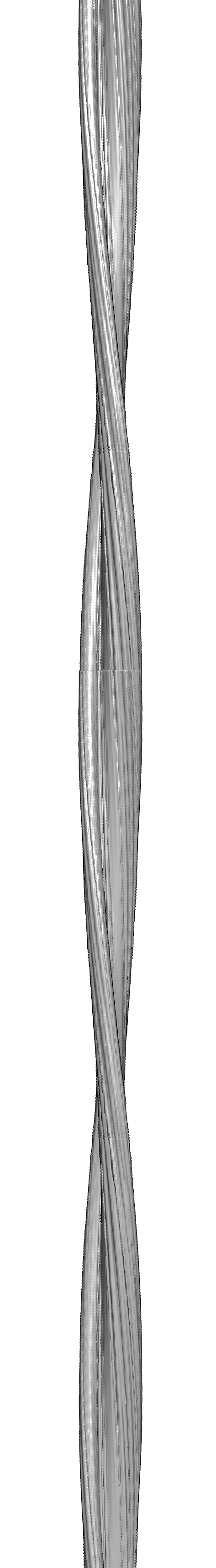 emd_3132.png emd_3132.png | 413.9 KB | ||
| Others | emd_3132_additional_1.map.gz | 14.1 MB | ||
| Archive directory |  http://ftp.pdbj.org/pub/emdb/structures/EMD-3132ftp://ftp.pdbj.org/pub/emdb/structures/EMD-3132 http://ftp.pdbj.org/pub/emdb/structures/EMD-3132ftp://ftp.pdbj.org/pub/emdb/structures/EMD-3132 | HTTPS FTP |
-Related structure data
| Related structure data |  5aefMC M: atomic model generated by this map C: citing same article ( |
|---|---|
| Similar structure data |
-Links
| EMDB pages | EMDB (EBI/PDBe) / EMDataResource |
|---|---|
| Related items in Molecule of the Month |


-Map
| File | Download / File: emd_3132.map.gz / Format: CCP4 / Size: 14.9 MB / Type: IMAGE STORED AS FLOATING POINT NUMBER (4 BYTES) | ||||||||||||||||||||||||||||||||||||||||||||||||||||||||||||
|---|---|---|---|---|---|---|---|---|---|---|---|---|---|---|---|---|---|---|---|---|---|---|---|---|---|---|---|---|---|---|---|---|---|---|---|---|---|---|---|---|---|---|---|---|---|---|---|---|---|---|---|---|---|---|---|---|---|---|---|---|---|
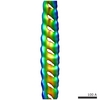
| Annotation | Reconstruction of an Abeta(1-42) amyloid-like fibril | ||||||||||||||||||||||||||||||||||||||||||||||||||||||||||||
| Projections & slices | Image control
Images are generated by Spider. generated in cubic-lattice coordinate | ||||||||||||||||||||||||||||||||||||||||||||||||||||||||||||
| Voxel size | X=Y=Z: 1.2 Å | ||||||||||||||||||||||||||||||||||||||||||||||||||||||||||||
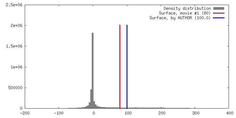
| Density |
| ||||||||||||||||||||||||||||||||||||||||||||||||||||||||||||
| Symmetry | Space group: 1 | ||||||||||||||||||||||||||||||||||||||||||||||||||||||||||||
| Details | EMDB XML:
CCP4 map header:
| ||||||||||||||||||||||||||||||||||||||||||||||||||||||||||||
 Z (Sec.)
 Y (Row.)
 X (Col.)


-Supplemental data
-Supplemental map: emd 3132 additional 1.map
| File | emd_3132_additional_1.map | ||||||||||||
|---|---|---|---|---|---|---|---|---|---|---|---|---|---|
| Projections & Slices |
| ||||||||||||
| Density Histograms |


- Sample components
-Entire : Abeta(1-42) amyloid-like fibril
| Entire | Name: Abeta(1-42) amyloid-like fibril |
|---|---|
| Components |
|
-Supramolecule #1000: Abeta(1-42) amyloid-like fibril
| Supramolecule | Name: Abeta(1-42) amyloid-like fibril / type: sample / ID: 1000 / Details: fibril / Oligomeric state: Peptide forms a helical assembly / Number unique components: 1 |
|---|
-Macromolecule #1: Abeta(1-42)
| Macromolecule | Name: Abeta(1-42) / type: protein_or_peptide / ID: 1 / Oligomeric state: Multimer / Recombinant expression: No / Database: NCBI |
|---|---|
| Source (natural) | Organism: Homo sapiens (human) / synonym: Human / Tissue: Brain |
| Molecular weight | Experimental: 4.418 KDa / Theoretical: 4.418 KDa |
-Experimental details
-Structure determination
| Method | cryo EM |
|---|---|
 Processing Processing | helical reconstruction |
| Aggregation state | filament |
-Sample preparation
| Concentration | 1 mg/mL |
|---|---|
| Buffer | pH: 7.4 / Details: 50mM Tris-HCl |
| Vitrification | Cryogen name: ETHANE / Chamber humidity: 40 % / Chamber temperature: 100 K / Instrument: GATAN CRYOPLUNGE 3 Method: grids: glow-discharged holey-carbon Blotting: 4 second backside blotting |
- Electron microscopy
| Microscope | FEI TECNAI F30 |
|---|---|
| Temperature | Average: 100 K |
| Date | Feb 5, 2010 |
| Image recording | Category: FILM / Film or detector model: KODAK SO-163 FILM / Digitization - Scanner: ZEISS SCAI / Digitization - Sampling interval: 7 µm / Number real images: 29 / Average electron dose: 30 e/Å2 / Bits/pixel: 14 |
| Tilt angle min | 0 |
| Tilt angle max | 0 |
| Electron beam | Acceleration voltage: 300 kV / Electron source:  FIELD EMISSION GUN FIELD EMISSION GUN |
| Electron optics | Calibrated magnification: 58333 / Illumination mode: FLOOD BEAM / Imaging mode: BRIGHT FIELD / Cs: 2.0 mm / Nominal defocus max: 3.0 µm / Nominal defocus min: 1.75 µm / Nominal magnification: 59000 |
| Sample stage | Specimen holder model: GATAN LIQUID NITROGEN |
| Experimental equipment |  Model: Tecnai F30 / Image courtesy: FEI Company |
-Image processing
| Details | Reconstruction was done with FREALIX. |
|---|---|
| Final reconstruction | Applied symmetry - Helical parameters - Δz: 4.7 Å Applied symmetry - Helical parameters - Δ&Phi: 0.769 ° Applied symmetry - Helical parameters - Axial symmetry: C2 (2 fold cyclic) Algorithm: OTHER / Resolution.type: BY AUTHOR / Resolution: 7.0 Å / Resolution method: OTHER / Software - Name: FREALIX Details: Final map was calculated from 29 single filament reconstructions. Resolution of data used during refinement never exceeded 10 angstrom resolution. Resolution of fibril core is about 5 angstroms. |
| CTF correction | Details: For each helical subunit |
| FSC plot (resolution estimation) |  |
