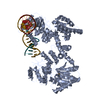
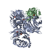
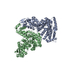
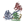
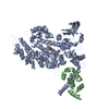
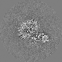
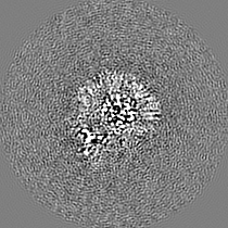
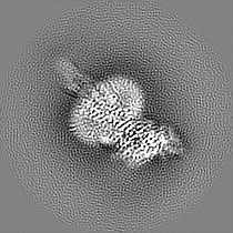
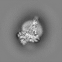
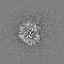
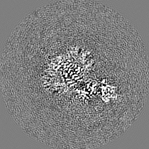
Journal: Biochem Biophys Res Commun / Year: 2020 Title: Structure of the human secretin receptor coupled to an engineered heterotrimeric G protein. Authors: Satoshi Fukuhara / Kazuhiro Kobayashi / Tsukasa Kusakizako / Wataru Iida / Masahiko Kato / Wataru Shihoya / Osamu Nureki / Abstract: Secretin is a gastrointestinal hormone that exerts multiple physiological functions via activation of the secretin receptor (SECR). SECR belongs to the class B G-protein-coupled receptors and is ...Secretin is a gastrointestinal hormone that exerts multiple physiological functions via activation of the secretin receptor (SECR). SECR belongs to the class B G-protein-coupled receptors and is involved in various processes, such as regulation of the pH of the duodenal content, food intake, and water homeostasis. Here, we report a cryo-electron microscopy structure of human SECR bound to secretin and an engineered Gs heterotrimer. The structure revealed the basic architecture of SECR and the secretin binding mode. A structural comparison of the SECR and PAC1R transmembrane domains revealed that transmembrane helices 1 and 2 play a prominent role in secretin recognition. Moreover, the extracellular domain of SECR is perpendicular to the TMD, unlike that of PAC1R. This comparison revealed the diverged peptide recognition mechanisms of these receptors, which belong to the same subgroup. Our structural information will facilitate drug discovery research for clinical applications.
History
Deposition
Sep 20, 2020
-
Header (metadata) release
Nov 4, 2020
-
Map release
Nov 4, 2020
-
Update
Nov 13, 2024
-
Current status
Nov 13, 2024
Processing site: PDBj / Status: Released
-
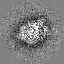
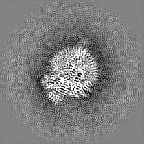
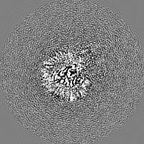
Structure visualization
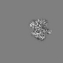
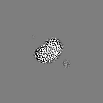
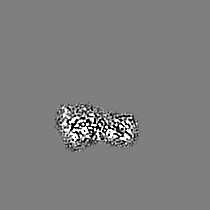
Movie
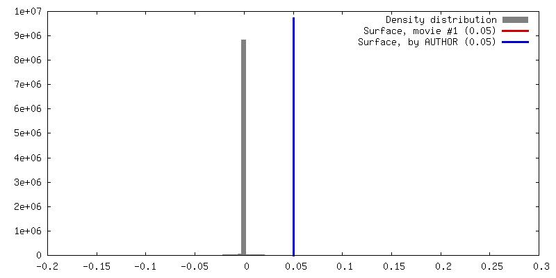
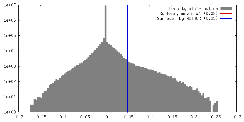
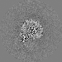
Surface view with section colored by density value
In the structure databanks used in Yorodumi, some data are registered as the other names, "COVID-19 virus" and "2019-nCoV". Here are the details of the virus and the list of structure data.
Jan 31, 2019. EMDB accession codes are about to change! (news from PDBe EMDB page)
EMDB accession codes are about to change! (news from PDBe EMDB page)
The allocation of 4 digits for EMDB accession codes will soon come to an end. Whilst these codes will remain in use, new EMDB accession codes will include an additional digit and will expand incrementally as the available range of codes is exhausted. The current 4-digit format prefixed with “EMD-” (i.e. EMD-XXXX) will advance to a 5-digit format (i.e. EMD-XXXXX), and so on. It is currently estimated that the 4-digit codes will be depleted around Spring 2019, at which point the 5-digit format will come into force.
The EM Navigator/Yorodumi systems omit the EMD- prefix.
Related info.:Q: What is EMD? / ID/Accession-code notation in Yorodumi/EM Navigator
Yorodumi is a browser for structure data from EMDB, PDB, SASBDB, etc.
This page is also the successor to EM Navigator detail page, and also detail information page/front-end page for Omokage search.
The word "yorodu" (or yorozu) is an old Japanese word meaning "ten thousand". "mi" (miru) is to see.
Related info.:EMDB / PDB / SASBDB / Comparison of 3 databanks / Yorodumi Search / Aug 31, 2016. New EM Navigator & Yorodumi / Yorodumi Papers / Jmol/JSmol / Function and homology information / Changes in new EM Navigator and Yorodumi
 Movie
Movie Controller


 Open data
 Basic information
 Map data
 Sample
 Keywords
 Function and homology information
 Homo sapiens (human) /
 Authors
 Citation
 Structure visualization
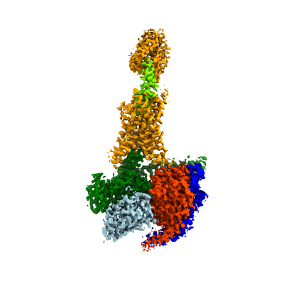
 Downloads & links
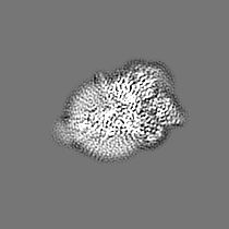
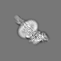
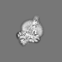
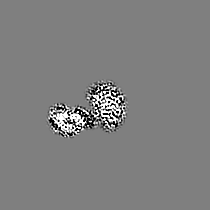
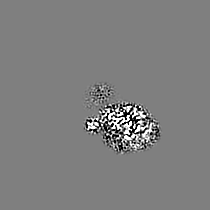
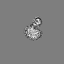
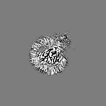
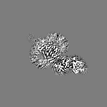
 emd_30566.png
 http://ftp.pdbj.org/pub/emdb/structures/EMD-30566


 Z (Sec.)
 Y (Row.)
 X (Col.)


 Sample components
 Spodoptera frugiperda (fall armyworm)
 Processing
 Electron microscopy
 FIELD EMISSION GUN