+ Open data
- Basic information
| Entry | Database: EMDB / ID: EMD-30322 | |||||||||
|---|---|---|---|---|---|---|---|---|---|---|
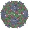
| Title | Coxsackievirus B1 F-particle | |||||||||
 Map data Map data | ||||||||||
 Sample Sample |
| |||||||||
 Keywords Keywords | Echovirus B / mature / VIRUS | |||||||||
| Function / homology |  Function and homology information Function and homology informationsymbiont-mediated suppression of host cytoplasmic pattern recognition receptor signaling pathway via inhibition of RIG-I activity / picornain 2A / symbiont-mediated suppression of host mRNA export from nucleus / symbiont genome entry into host cell via pore formation in plasma membrane / picornain 3C / T=pseudo3 icosahedral viral capsid / host cell cytoplasmic vesicle membrane / endocytosis involved in viral entry into host cell / nucleoside-triphosphate phosphatase / channel activity ...symbiont-mediated suppression of host cytoplasmic pattern recognition receptor signaling pathway via inhibition of RIG-I activity / picornain 2A / symbiont-mediated suppression of host mRNA export from nucleus / symbiont genome entry into host cell via pore formation in plasma membrane / picornain 3C / T=pseudo3 icosahedral viral capsid / host cell cytoplasmic vesicle membrane / endocytosis involved in viral entry into host cell / nucleoside-triphosphate phosphatase / channel activity / viral capsid / monoatomic ion transmembrane transport / host cell cytoplasm / DNA replication / RNA helicase activity / induction by virus of host autophagy / RNA-directed RNA polymerase / viral RNA genome replication / cysteine-type endopeptidase activity / RNA-dependent RNA polymerase activity / virus-mediated perturbation of host defense response / DNA-templated transcription / host cell nucleus / virion attachment to host cell / structural molecule activity / ATP hydrolysis activity / proteolysis / RNA binding / ATP binding / membrane / metal ion binding / cytoplasm Similarity search - Function | |||||||||
| Biological species |  Coxsackievirus B1 Coxsackievirus B1 | |||||||||
| Method | single particle reconstruction / cryo EM / Resolution: 3.6 Å | |||||||||
 Authors Authors | Feng R / Wang K | |||||||||
| Funding support |  China, 1 items China, 1 items
| |||||||||
 Citation Citation |  Journal: Nat Commun / Year: 2020 Journal: Nat Commun / Year: 2020Title: Structures of Echovirus 30 in complex with its receptors inform a rational prediction for enterovirus receptor usage. Authors: Kang Wang / Ling Zhu / Yao Sun / Minhao Li / Xin Zhao / Lunbiao Cui / Li Zhang / George F Gao / Weiwei Zhai / Fengcai Zhu / Zihe Rao / Xiangxi Wang /  Abstract: Receptor usage that determines cell tropism and drives viral classification closely correlates with the virus structure. Enterovirus B (EV-B) consists of several subgroups according to receptor ...Receptor usage that determines cell tropism and drives viral classification closely correlates with the virus structure. Enterovirus B (EV-B) consists of several subgroups according to receptor usage, among which echovirus 30 (E30), a leading causative agent for human aseptic meningitis, utilizes FcRn as an uncoating receptor. However, receptors for many EVs remain unknown. Here we analyzed the atomic structures of E30 mature virion, empty- and A-particles, which reveals serotype-specific epitopes and striking conformational differences between the subgroups within EV-Bs. Of these, the VP1 BC loop markedly distinguishes E30 from other EV-Bs, indicative of a role as a structural marker for EV-B. By obtaining cryo-electron microscopy structures of E30 in complex with its receptor FcRn and CD55 and comparing its homologs, we deciphered the underlying molecular basis for receptor recognition. Together with experimentally derived viral receptor identifications, we developed a structure-based in silico algorithm to inform a rational prediction for EV receptor usage. | |||||||||
| History |
|
- Structure visualization
| Movie |
 Movie viewer Movie viewer |
|---|---|
| Structure viewer | EM map:  SurfView SurfView Molmil Molmil Jmol/JSmol Jmol/JSmol |
| Supplemental images |
- Downloads & links
-EMDB archive
| Map data |  emd_30322.map.gz emd_30322.map.gz | 116.7 MB |  EMDB map data format EMDB map data format | |
|---|---|---|---|---|
| Header (meta data) |  emd-30322-v30.xml emd-30322-v30.xml emd-30322.xml emd-30322.xml | 14.9 KB 14.9 KB | Display Display |  EMDB header EMDB header |
| Images |  emd_30322.png emd_30322.png | 126.4 KB | ||
| Filedesc metadata |  emd-30322.cif.gz emd-30322.cif.gz | 5.9 KB | ||
| Archive directory |  http://ftp.pdbj.org/pub/emdb/structures/EMD-30322 http://ftp.pdbj.org/pub/emdb/structures/EMD-30322 ftp://ftp.pdbj.org/pub/emdb/structures/EMD-30322 ftp://ftp.pdbj.org/pub/emdb/structures/EMD-30322 | HTTPS FTP |
-Validation report
| Summary document |  emd_30322_validation.pdf.gz emd_30322_validation.pdf.gz | 805.8 KB | Display |  EMDB validaton report EMDB validaton report |
|---|---|---|---|---|
| Full document |  emd_30322_full_validation.pdf.gz emd_30322_full_validation.pdf.gz | 805.4 KB | Display | |
| Data in XML |  emd_30322_validation.xml.gz emd_30322_validation.xml.gz | 6.5 KB | Display | |
| Data in CIF |  emd_30322_validation.cif.gz emd_30322_validation.cif.gz | 7.4 KB | Display | |
| Arichive directory |  https://ftp.pdbj.org/pub/emdb/validation_reports/EMD-30322 https://ftp.pdbj.org/pub/emdb/validation_reports/EMD-30322 ftp://ftp.pdbj.org/pub/emdb/validation_reports/EMD-30322 ftp://ftp.pdbj.org/pub/emdb/validation_reports/EMD-30322 | HTTPS FTP |
-Related structure data
| Related structure data |  7c9zMC  7c9sC  7c9tC  7c9uC  7c9vC  7c9wC  7c9xC  7c9yC M: atomic model generated by this map C: citing same article ( |
|---|---|
| Similar structure data |
- Links
Links
| EMDB pages |  EMDB (EBI/PDBe) / EMDB (EBI/PDBe) /  EMDataResource EMDataResource |
|---|---|
| Related items in Molecule of the Month |
- Map
Map
| File |  Download / File: emd_30322.map.gz / Format: CCP4 / Size: 125 MB / Type: IMAGE STORED AS FLOATING POINT NUMBER (4 BYTES) Download / File: emd_30322.map.gz / Format: CCP4 / Size: 125 MB / Type: IMAGE STORED AS FLOATING POINT NUMBER (4 BYTES) | ||||||||||||||||||||||||||||||||||||||||||||||||||||||||||||
|---|---|---|---|---|---|---|---|---|---|---|---|---|---|---|---|---|---|---|---|---|---|---|---|---|---|---|---|---|---|---|---|---|---|---|---|---|---|---|---|---|---|---|---|---|---|---|---|---|---|---|---|---|---|---|---|---|---|---|---|---|---|
| Projections & slices | Image control
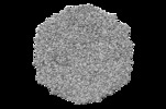
Images are generated by Spider. | ||||||||||||||||||||||||||||||||||||||||||||||||||||||||||||
| Voxel size | X=Y=Z: 1.345 Å | ||||||||||||||||||||||||||||||||||||||||||||||||||||||||||||
| Density |
| ||||||||||||||||||||||||||||||||||||||||||||||||||||||||||||
| Symmetry | Space group: 1 | ||||||||||||||||||||||||||||||||||||||||||||||||||||||||||||
| Details | EMDB XML:
CCP4 map header:
| ||||||||||||||||||||||||||||||||||||||||||||||||||||||||||||
-Supplemental data
- Sample components
-Entire : Coxsackievirus B1
| Entire | Name:  Coxsackievirus B1 Coxsackievirus B1 |
|---|---|
| Components |
|
-Supramolecule #1: Coxsackievirus B1
| Supramolecule | Name: Coxsackievirus B1 / type: virus / ID: 1 / Parent: 0 / Macromolecule list: #1-#4 Details: Particles purified from the cell cultures innoculated with the live CB1. NCBI-ID: 12071 / Sci species name: Coxsackievirus B1 / Virus type: VIRION / Virus isolate: SEROTYPE / Virus enveloped: No / Virus empty: No |
|---|---|
| Host (natural) | Organism:  Homo sapiens (human) Homo sapiens (human) |
| Virus shell | Shell ID: 1 / Diameter: 30.0 Å / T number (triangulation number): 3 |
-Macromolecule #1: VP1
| Macromolecule | Name: VP1 / type: protein_or_peptide / ID: 1 / Number of copies: 1 / Enantiomer: LEVO |
|---|---|
| Source (natural) | Organism:  Coxsackievirus B1 Coxsackievirus B1 |
| Molecular weight | Theoretical: 31.247 KDa |
| Sequence | String: GPVEESVERA MVRVADTVSS KPTNSESIPA LTAAETGHTS QVVPSDTMQT RHVKNYHSRS ESSIENFLCR SACVYYATYN NNSEKGYAE WVINTRQVAQ LLRRKLEFTY LRFDLELTFV ITSAQEPSTA TSVDAPVQTQ QIMYVPPGGP VPTKVTDYAW Q TSTNPSVF ...String: GPVEESVERA MVRVADTVSS KPTNSESIPA LTAAETGHTS QVVPSDTMQT RHVKNYHSRS ESSIENFLCR SACVYYATYN NNSEKGYAE WVINTRQVAQ LLRRKLEFTY LRFDLELTFV ITSAQEPSTA TSVDAPVQTQ QIMYVPPGGP VPTKVTDYAW Q TSTNPSVF WTEGNAPPRM SIPFISIGNA YSCFYDGWTQ FSRNGVYGIN TLNNMGTLYM RHVNEAGQGP IKSTVRIYFK PK HVKAWVP RPPRLCQYEK QKNVNFNPTG VTTTRSNITT T UniProtKB: Genome polyprotein |
-Macromolecule #2: VP2
| Macromolecule | Name: VP2 / type: protein_or_peptide / ID: 2 / Number of copies: 1 / Enantiomer: LEVO |
|---|---|
| Source (natural) | Organism:  Coxsackievirus B1 Coxsackievirus B1 |
| Molecular weight | Theoretical: 29.125768 KDa |
| Sequence | String: SPSAEECGYS DRVRSITLGN STITTQECAN VVVGYGVWPE YLKDNEATGE DQPTQPDVAT CRFYTLESVQ WMKNSAGWWW KLPDALSQM GLFGQNMQYH YLGRTGYTIH VQCNASKFHQ GCLLVVCVPE AEMGCSNLNN TPKFAELSGG DNARMFTDTE V GTSNDKKV ...String: SPSAEECGYS DRVRSITLGN STITTQECAN VVVGYGVWPE YLKDNEATGE DQPTQPDVAT CRFYTLESVQ WMKNSAGWWW KLPDALSQM GLFGQNMQYH YLGRTGYTIH VQCNASKFHQ GCLLVVCVPE AEMGCSNLNN TPKFAELSGG DNARMFTDTE V GTSNDKKV QTAVWNAGMG VGVGNLTIFP HQWINLRTNN SATIVMPYIN SVPMDNMYRH NNLTLMIIPF VPLNYSEGSS PY VPITVTI APMCAEYNGL RLASSQ UniProtKB: Genome polyprotein |
-Macromolecule #3: VP3
| Macromolecule | Name: VP3 / type: protein_or_peptide / ID: 3 / Number of copies: 1 / Enantiomer: LEVO |
|---|---|
| Source (natural) | Organism:  Coxsackievirus B1 Coxsackievirus B1 |
| Molecular weight | Theoretical: 26.507973 KDa |
| Sequence | String: GLPVMTTPGS TQFLTSDDFQ SPSAMPQFDV TPEMQIPGRV NNLMEIAEVD SVVPVNNTDN NVNGLKAYQI PVQSNSDNRR QVFGFPLQP GANNVLNRTL LGEILNYYTH WSGSIKLTFM FCGSAMATGK FLLAYSPPGA GVPKNRRDAM LGTHVIWDVG L QSSCVLCV ...String: GLPVMTTPGS TQFLTSDDFQ SPSAMPQFDV TPEMQIPGRV NNLMEIAEVD SVVPVNNTDN NVNGLKAYQI PVQSNSDNRR QVFGFPLQP GANNVLNRTL LGEILNYYTH WSGSIKLTFM FCGSAMATGK FLLAYSPPGA GVPKNRRDAM LGTHVIWDVG L QSSCVLCV PWISQTHYRY VVEDEYTAAG YVTCWYQTNI IVPADVQSTC DILCFVSACN DFSVRMLKDT PFIRQDNFYQ UniProtKB: Genome polyprotein |
-Macromolecule #4: VP4
| Macromolecule | Name: VP4 / type: protein_or_peptide / ID: 4 / Number of copies: 1 / Enantiomer: LEVO |
|---|---|
| Source (natural) | Organism:  Coxsackievirus B1 Coxsackievirus B1 |
| Molecular weight | Theoretical: 7.367077 KDa |
| Sequence | String: GAQVSTQKTG AHETGLNASG NSIIHYTNIN YYKDAASNSA NRQDFTQDPG KFTEPVKDIM IKSMPALN UniProtKB: VP4 |
-Macromolecule #5: PALMITIC ACID
| Macromolecule | Name: PALMITIC ACID / type: ligand / ID: 5 / Number of copies: 1 / Formula: PLM |
|---|---|
| Molecular weight | Theoretical: 256.424 Da |
| Chemical component information |  ChemComp-PLM: |
-Macromolecule #6: MYRISTIC ACID
| Macromolecule | Name: MYRISTIC ACID / type: ligand / ID: 6 / Number of copies: 1 / Formula: MYR |
|---|---|
| Molecular weight | Theoretical: 228.371 Da |
| Chemical component information |  ChemComp-MYR: |
-Experimental details
-Structure determination
| Method | cryo EM |
|---|---|
 Processing Processing | single particle reconstruction |
| Aggregation state | particle |
- Sample preparation
| Buffer | pH: 7.4 |
|---|---|
| Grid | Model: Quantifoil R1.2/1.3 / Material: GOLD / Support film - Material: CARBON / Support film - topology: HOLEY |
| Vitrification | Cryogen name: ETHANE / Chamber humidity: 100 % / Instrument: FEI VITROBOT MARK I |
- Electron microscopy
| Microscope | FEI TITAN KRIOS |
|---|---|
| Image recording | Film or detector model: GATAN K2 SUMMIT (4k x 4k) / Detector mode: SUPER-RESOLUTION / Average electron dose: 30.0 e/Å2 |
| Electron beam | Acceleration voltage: 300 kV / Electron source:  FIELD EMISSION GUN FIELD EMISSION GUN |
| Electron optics | Illumination mode: FLOOD BEAM / Imaging mode: DARK FIELD / Cs: 2.7 mm |
| Sample stage | Specimen holder model: FEI TITAN KRIOS AUTOGRID HOLDER / Cooling holder cryogen: NITROGEN |
| Experimental equipment |  Model: Titan Krios / Image courtesy: FEI Company |
- Image processing
| Startup model | Type of model: INSILICO MODEL |
|---|---|
| Final reconstruction | Applied symmetry - Point group: I (icosahedral) / Resolution.type: BY AUTHOR / Resolution: 3.6 Å / Resolution method: FSC 0.143 CUT-OFF / Software - Name: RELION (ver. 3.0) / Number images used: 5568 |
| Initial angle assignment | Type: PROJECTION MATCHING |
| Final angle assignment | Type: PROJECTION MATCHING |
-Atomic model buiding 1
| Refinement | Space: REAL / Protocol: RIGID BODY FIT |
|---|---|
| Output model |  PDB-7c9z: |
 Movie
Movie Controller


 Z (Sec.)
 Y (Row.)
 X (Col.)


