National Natural Science Foundation of China (NSFC)
China
Citation
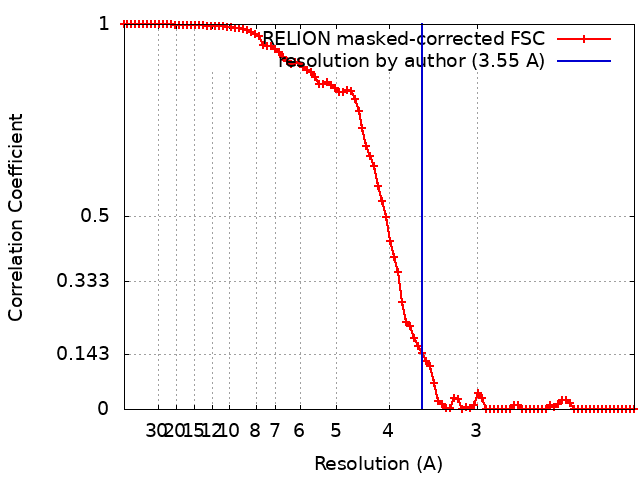
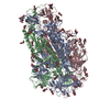
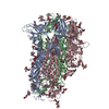
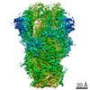
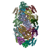
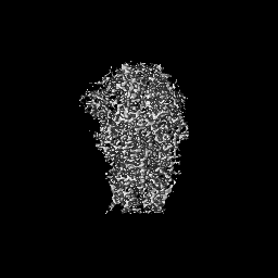
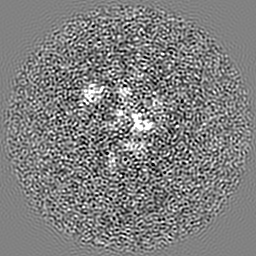
Journal: J Virol / Year: 2020 Title: Cryo-electron Microscopy Structure of the Swine Acute Diarrhea Syndrome Coronavirus Spike Glycoprotein Provides Insights into Evolution of Unique Coronavirus Spike Proteins. Authors: Hongxin Guan / Youwang Wang / Vanja Perčulija / Abdullah F U H Saeed / Yichang Liu / Jinyu Li / Syed Sajid Jan / Yu Li / Ping Zhu / Songying Ouyang / Abstract: Coronaviruses (CoV) have caused a number of major epidemics in humans and animals, including the current pandemic of coronavirus disease 2019 (COVID-19), which has brought a renewed focus on the ...Coronaviruses (CoV) have caused a number of major epidemics in humans and animals, including the current pandemic of coronavirus disease 2019 (COVID-19), which has brought a renewed focus on the evolution and interspecies transmission of coronaviruses. Swine acute diarrhea syndrome coronavirus (SADS-CoV), which was recently identified in piglets in southern China, is an alphacoronavirus that originates from the same genus of horseshoe bats as severe acute respiratory syndrome CoV (SARS-CoV) and that was reported to be capable of infecting cells from a broad range of species, suggesting a considerable potential for interspecies transmission. Given the importance of the coronavirus spike (S) glycoprotein in host range determination and viral entry, we report a cryo-electron microscopy (cryo-EM) structure of the SADS-CoV S trimer in the prefusion conformation at a 3.55-Å resolution. Our structure reveals that the SADS-CoV S trimer assumes an intrasubunit quaternary packing mode in which the S1 subunit N-terminal domain (S1-NTD) and the S1 subunit C-terminal domain (S1-CTD) of the same protomer pack together by facing each other in the lying-down state. SADS-CoV S has several distinctive structural features that may facilitate immune escape, such as a relatively compact architecture of the S trimer and epitope masking by glycan shielding. Comparison of SADS-CoV S with the spike proteins of the other coronavirus genera suggested that the structural features of SADS-CoV S are evolutionarily related to those of the spike proteins of the other genera rather than to the spike protein of a typical alphacoronavirus. These data provide new insights into the evolutionary relationship between spike glycoproteins of SADS-CoV and those of other coronaviruses and extend our understanding of their structural and functional diversity. In this article, we report the atomic-resolution prefusion structure of the spike protein from swine acute diarrhea syndrome coronavirus (SADS-CoV). SADS-CoV is a pathogenic alphacoronavirus that was responsible for a large-scale outbreak of fatal disease in pigs and that was reported to be capable of interspecies transmission. We describe the overall structure of the SADS-CoV spike protein and conducted a detailed analysis of its main structural elements. Our results and analyses are consistent with those of previous phylogenetic studies and suggest that the SADS-CoV spike protein is evolutionarily related to the spike proteins of betacoronaviruses, with a strong similarity in S1-NTDs and a marked divergence in S1-CTDs. Moreover, we discuss the possible immune evasion strategies used by the SADS-CoV spike protein. Our study provides insights into the structure and immune evasion strategies of the SADS-CoV spike protein and broadens the understanding of the evolutionary relationships between coronavirus spike proteins of different genera.
History
Deposition
Mar 3, 2020
-
Header (metadata) release
Aug 26, 2020
-
Map release
Aug 26, 2020
-
Update
Nov 20, 2024
-
Current status
Nov 20, 2024
Processing site: PDBj / Status: Released
-
Structure visualization
Movie
Surface view with section colored by density value
In the structure databanks used in Yorodumi, some data are registered as the other names, "COVID-19 virus" and "2019-nCoV". Here are the details of the virus and the list of structure data.
Jan 31, 2019. EMDB accession codes are about to change! (news from PDBe EMDB page)
EMDB accession codes are about to change! (news from PDBe EMDB page)
The allocation of 4 digits for EMDB accession codes will soon come to an end. Whilst these codes will remain in use, new EMDB accession codes will include an additional digit and will expand incrementally as the available range of codes is exhausted. The current 4-digit format prefixed with “EMD-” (i.e. EMD-XXXX) will advance to a 5-digit format (i.e. EMD-XXXXX), and so on. It is currently estimated that the 4-digit codes will be depleted around Spring 2019, at which point the 5-digit format will come into force.
The EM Navigator/Yorodumi systems omit the EMD- prefix.
Related info.:Q: What is EMD? / ID/Accession-code notation in Yorodumi/EM Navigator
Yorodumi is a browser for structure data from EMDB, PDB, SASBDB, etc.
This page is also the successor to EM Navigator detail page, and also detail information page/front-end page for Omokage search.
The word "yorodu" (or yorozu) is an old Japanese word meaning "ten thousand". "mi" (miru) is to see.
Related info.:EMDB / PDB / SASBDB / Comparison of 3 databanks / Yorodumi Search / Aug 31, 2016. New EM Navigator & Yorodumi / Yorodumi Papers / Jmol/JSmol / Function and homology information / Changes in new EM Navigator and Yorodumi
 Movie
Movie Controller


 Open data
 Basic information
 Map data
 Sample
 Keywords
 Function and homology information
 Swine acute diarrhea syndrome coronavirus
 Authors
 China, 1 items
  Citation
 Structure visualization
 Downloads & links
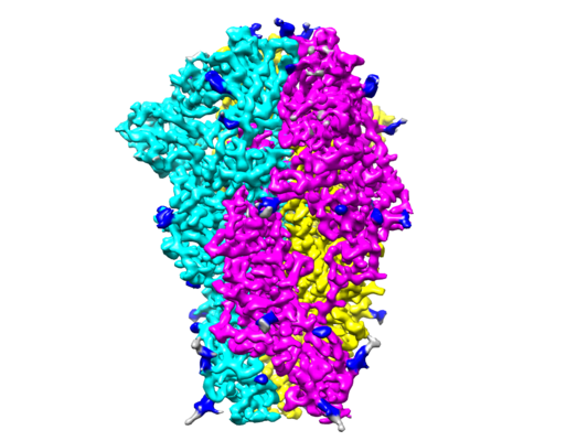 emd_30071.png
 http://ftp.pdbj.org/pub/emdb/structures/EMD-30071

 Z (Sec.)
 Y (Row.)
 X (Col.)


 Sample components
 Processing
 Electron microscopy
 FIELD EMISSION GUN