[English] 日本語
 Yorodumi
- PDB-2y9j: THREE-DIMENSIONAL MODEL OF SALMONELLA'S NEEDLE COMPLEX AT SUBNANO... -
+ Open data
- Basic information
| Entry | Database: PDB / ID: 2y9j | ||||||
|---|---|---|---|---|---|---|---|
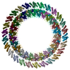
| Title | THREE-DIMENSIONAL MODEL OF SALMONELLA'S NEEDLE COMPLEX AT SUBNANOMETER RESOLUTION | ||||||
 Components Components |
| ||||||
 Keywords Keywords | PROTEIN TRANSPORT / TYPE III SECRETION / IR1 / INNER MEMBRANE RING / C24-FOLD | ||||||
| Function / homology |  Function and homology information Function and homology information | ||||||
| Biological species |  SALMONELLA ENTERICA SUBSP. ENTERICA SEROVAR TYPHIMURIUM (bacteria) SALMONELLA ENTERICA SUBSP. ENTERICA SEROVAR TYPHIMURIUM (bacteria) | ||||||
| Method | ELECTRON MICROSCOPY / single particle reconstruction / cryo EM / Resolution: 6.4 Å | ||||||
 Authors Authors | Schraidt, O. / Marlovits, T.C. | ||||||
 Citation Citation |  Journal: Science / Year: 2011 Journal: Science / Year: 2011Title: Three-dimensional model of Salmonella's needle complex at subnanometer resolution. Authors: Oliver Schraidt / Thomas C Marlovits /  Abstract: Type III secretion systems (T3SSs) are essential virulence factors used by many Gram-negative bacteria to inject proteins that make eukaryotic host cells accessible to invasion. The T3SS core ...Type III secretion systems (T3SSs) are essential virulence factors used by many Gram-negative bacteria to inject proteins that make eukaryotic host cells accessible to invasion. The T3SS core structure, the needle complex (NC), is a ~3.5 megadalton-sized, oligomeric, membrane-embedded complex. Analyzing cryo-electron microscopy images of top views of NCs or NC substructures from Salmonella typhimurium revealed a 24-fold symmetry for the inner rings and a 15-fold symmetry for the outer rings, giving an overall C3 symmetry. Local refinement and averaging showed the organization of the central core and allowed us to reconstruct a subnanometer composite structure of the NC, which together with confident docking of atomic structures reveal insights into its overall organization and structural requirements during assembly. | ||||||
| History |
|
- Structure visualization
| Movie |
 Movie viewer Movie viewer |
|---|---|
| Structure viewer | Molecule:  Molmil Molmil Jmol/JSmol Jmol/JSmol |
- Downloads & links
- Download
| PDBx/mmCIF format |  2y9j.cif.gz 2y9j.cif.gz | 1.5 MB | Display |  PDBx/mmCIF format PDBx/mmCIF format |
|---|---|---|---|---|
| PDB format |  pdb2y9j.ent.gz pdb2y9j.ent.gz | 1.3 MB | Display |  PDB format PDB format |
| PDBx/mmJSON format |  2y9j.json.gz 2y9j.json.gz | Tree view |  PDBx/mmJSON format PDBx/mmJSON format | |
| Others |  Other downloads Other downloads |
-Validation report
| Arichive directory |  https://data.pdbj.org/pub/pdb/validation_reports/y9/2y9j https://data.pdbj.org/pub/pdb/validation_reports/y9/2y9j ftp://data.pdbj.org/pub/pdb/validation_reports/y9/2y9j ftp://data.pdbj.org/pub/pdb/validation_reports/y9/2y9j | HTTPS FTP |
|---|
-Related structure data
| Related structure data |  1874MC  1871C  1875C  2y9kC C: citing same article ( M: map data used to model this data |
|---|---|
| Similar structure data |
- Links
Links
- Assembly
| Deposited unit | 
|
|---|---|
| 1 |
|
- Components
| #1: Protein | Mass: 21839.814 Da / Num. of mol.: 24 / Fragment: PERIPLASMIC DOMAIN, RESIDUES 177-362 / Source method: isolated from a natural source Source: (natural)  SALMONELLA ENTERICA SUBSP. ENTERICA SEROVAR TYPHIMURIUM (bacteria) SALMONELLA ENTERICA SUBSP. ENTERICA SEROVAR TYPHIMURIUM (bacteria)References: UniProt: P41783 #2: Protein | Mass: 19015.523 Da / Num. of mol.: 24 / Fragment: PERIPLASMIC DOMAIN, RESIDUES 21-190 / Source method: isolated from a natural source Source: (natural)  SALMONELLA ENTERICA SUBSP. ENTERICA SEROVAR TYPHIMURIUM (bacteria) SALMONELLA ENTERICA SUBSP. ENTERICA SEROVAR TYPHIMURIUM (bacteria)References: UniProt: P41786 #3: Water | ChemComp-HOH / | |
|---|
-Experimental details
-Experiment
| Experiment | Method: ELECTRON MICROSCOPY |
|---|---|
| EM experiment | Aggregation state: PARTICLE / 3D reconstruction method: single particle reconstruction |
- Sample preparation
| Component | Name: INNER MEMBRANE RING 1 / Type: COMPLEX |
|---|---|
| Buffer solution | pH: 7.5 / Details: 10mM Tris-HCl, 0.5M NaCl, 0.1% LDAO |
| Specimen | Embedding applied: NO / Shadowing applied: NO / Staining applied: NO / Vitrification applied: YES |
| Specimen support | Details: CARBON |
| Vitrification | Cryogen name: ETHANE / Details: LIQUID ETHANE |
- Electron microscopy imaging
| Experimental equipment |  Model: Tecnai Polara / Image courtesy: FEI Company |
|---|---|
| Microscopy | Model: FEI POLARA 300 Details: MAGNIFICATION AT THE CCD 112969, CAMERA PIXEL 15UM, 1.33 ANGSTROM PER PIXEL, DATA COLLECTED SEMI- AUTOMATICALLY WITH POINT-2-POINT (DEVELOPED IN-HOUSE) |
| Electron gun | Electron source:  FIELD EMISSION GUN / Accelerating voltage: 300 kV / Illumination mode: FLOOD BEAM FIELD EMISSION GUN / Accelerating voltage: 300 kV / Illumination mode: FLOOD BEAM |
| Electron lens | Mode: BRIGHT FIELD / Nominal magnification: 93000 X / Nominal defocus max: 2500 nm / Nominal defocus min: 1000 nm / Cs: 2 mm |
| Image recording | Film or detector model: GATAN ULTRASCAN 4000 (4k x 4k) |
| Radiation wavelength | Relative weight: 1 |
- Processing
| EM software |
| ||||||||||||
|---|---|---|---|---|---|---|---|---|---|---|---|---|---|
| CTF correction | Details: EACH MICROGRAPH | ||||||||||||
| Symmetry | Point symmetry: C24 (24 fold cyclic) | ||||||||||||
| 3D reconstruction | Method: PROJECTION MATCHING / Resolution: 6.4 Å / Resolution method: FSC 0.5 CUT-OFF / Actual pixel size: 1.33 Å Details: RESOLUTION 6.4 ANGSTROM (0.5 FSC), 4.7 ANGSTROM (HALF BIT) PRGH (3GR1) MONOMERS WERE FITTED SEPARATELY, PRGK MODEL WAS BUILT BASED ON HOMOLOGY MODEL OF ESCJ (1YJ7) AND THEN INDIVIDUALLY ...Details: RESOLUTION 6.4 ANGSTROM (0.5 FSC), 4.7 ANGSTROM (HALF BIT) PRGH (3GR1) MONOMERS WERE FITTED SEPARATELY, PRGK MODEL WAS BUILT BASED ON HOMOLOGY MODEL OF ESCJ (1YJ7) AND THEN INDIVIDUALLY FITTED INTO EM MAP. SUBMISSION BASED ON EXPERIMENTAL DATA FROM EMDB EMD-1874. (DEPOSITION ID: 7821). Symmetry type: POINT | ||||||||||||
| Atomic model building | Protocol: OTHER / Space: REAL / Target criteria: Cross-correlation coefficient / Details: METHOD--CORRELATION | ||||||||||||
| Atomic model building | PDB-ID: 2Y9J Accession code: 2Y9J / Source name: PDB / Type: experimental model | ||||||||||||
| Refinement | Highest resolution: 6.4 Å | ||||||||||||
| Refinement step | Cycle: LAST / Highest resolution: 6.4 Å
|
 Movie
Movie Controller


 PDBj
PDBj


