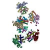
 Movie
Movie Controller

[English] 日本語
 Yorodumi
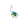
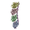
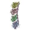
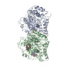
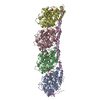
- PDB-2xrp: Human Doublecortin N-DC Repeat (1MJD) and Mammalian Tubulin (1JFF... -
+ Open data

- Basic information
| Entry | Database: PDB / ID: 2xrp | |||||||||
|---|---|---|---|---|---|---|---|---|---|---|
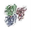
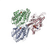
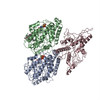
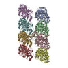
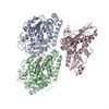
| Title | Human Doublecortin N-DC Repeat (1MJD) and Mammalian Tubulin (1JFF and 3HKE) Docked into the 8-Angstrom Cryo-EM Map of Doublecortin- Stabilised Microtubules | |||||||||
 Components Components |
| |||||||||
 Keywords Keywords | STRUCTURAL PROTEIN | |||||||||
| Function / homology |  Function and homology information Function and homology informationaxoneme assembly / Neurofascin interactions / positive regulation of axon guidance / microtubule associated complex / microtubule-based process / central nervous system development / structural constituent of cytoskeleton / microtubule cytoskeleton organization / neuron migration / nervous system development ...axoneme assembly / Neurofascin interactions / positive regulation of axon guidance / microtubule associated complex / microtubule-based process / central nervous system development / structural constituent of cytoskeleton / microtubule cytoskeleton organization / neuron migration / nervous system development / mitotic cell cycle / retina development in camera-type eye / microtubule cytoskeleton / microtubule binding / Hydrolases; Acting on acid anhydrides; Acting on GTP to facilitate cellular and subcellular movement / microtubule / cytoskeleton / neuron projection / hydrolase activity / intracellular signal transduction / protein heterodimerization activity / GTPase activity / protein kinase binding / GTP binding / metal ion binding / cytosol / cytoplasm Similarity search - Function | |||||||||
| Biological species |  HOMO SAPIENS (human) HOMO SAPIENS (human) | |||||||||
| Method | ELECTRON MICROSCOPY / single particle reconstruction / cryo EM / Resolution: 8.2 Å | |||||||||
 Authors Authors | Fourniol, F.J. / Sindelar, C.V. / Amigues, B. / Clare, D.K. / Thomas, G. / Perderiset, M. / Francis, F. / Houdusse, A. / Moores, C.A. | |||||||||
 Citation Citation | Journal: J Cell Biol / Year: 2010 Title: Template-free 13-protofilament microtubule-MAP assembly visualized at 8 A resolution. Authors: Franck J Fourniol / Charles V Sindelar / Béatrice Amigues / Daniel K Clare / Geraint Thomas / Mylène Perderiset / Fiona Francis / Anne Houdusse / Carolyn A Moores /  Abstract: Microtubule-associated proteins (MAPs) are essential for regulating and organizing cellular microtubules (MTs). However, our mechanistic understanding of MAP function is limited by a lack of detailed ...Microtubule-associated proteins (MAPs) are essential for regulating and organizing cellular microtubules (MTs). However, our mechanistic understanding of MAP function is limited by a lack of detailed structural information. Using cryo-electron microscopy and single particle algorithms, we solved the 8 Å structure of doublecortin (DCX)-stabilized MTs. Because of DCX's unusual ability to specifically nucleate and stabilize 13-protofilament MTs, our reconstruction provides unprecedented insight into the structure of MTs with an in vivo architecture, and in the absence of a stabilizing drug. DCX specifically recognizes the corner of four tubulin dimers, a binding mode ideally suited to stabilizing both lateral and longitudinal lattice contacts. A striking consequence of this is that DCX does not bind the MT seam. DCX binding on the MT surface indirectly stabilizes conserved tubulin-tubulin lateral contacts in the MT lumen, operating independently of the nucleotide bound to tubulin. DCX's exquisite binding selectivity uncovers important insights into regulation of cellular MTs. | |||||||||
| History |
|
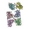
- Structure visualization
| Movie |
Movie viewer |
|---|---|
| Structure viewer | Molecule: MolmilJmol/JSmol |
- Downloads & links
-Download
| PDBx/mmCIF format | 2xrp.cif.gz | 682.7 KB | Display | PDBx/mmCIF format |
|---|---|---|---|---|
| PDB format | pdb2xrp.ent.gz | 545.1 KB | Display | PDB format |
| PDBx/mmJSON format | 2xrp.json.gz | Tree view | PDBx/mmJSON format | |
| Others |  Other downloads Other downloads |
-Validation report
| Summary document | 2xrp_validation.pdf.gz | 1.4 MB | Display | wwPDB validaton report |
|---|---|---|---|---|
| Full document | 2xrp_full_validation.pdf.gz | 1.8 MB | Display | |
| Data in XML | 2xrp_validation.xml.gz | 157.7 KB | Display | |
| Data in CIF | 2xrp_validation.cif.gz | 221.8 KB | Display | |
| Arichive directory | https://data.pdbj.org/pub/pdb/validation_reports/xr/2xrpftp://data.pdbj.org/pub/pdb/validation_reports/xr/2xrp | HTTPS FTP |
-Related structure data
| Related structure data | 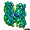 1788MC 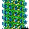 1787C M: map data used to model this data C: citing same article ( |
|---|---|
| Similar structure data |


-Links
 PDBj
PDBj


- Assembly
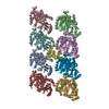
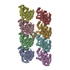
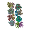
| Deposited unit | 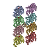
|
|---|---|
| 1 |
|
-Components
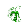
| #1: Protein | Mass: 49907.770 Da / Num. of mol.: 4 / Source method: isolated from a natural source / Source: (natural) #2: Protein | Mass: 50236.352 Da / Num. of mol.: 4 / Source method: isolated from a natural source / Source: (natural) #3: Protein | | Mass: 11029.393 Da / Num. of mol.: 1 / Fragment: RESIDUES 46-140 Source method: isolated from a genetically manipulated source Source: (gene. exp.) HOMO SAPIENS (human) / Organ: BRAIN / Plasmid: PFASTBAC / Production host:   SPODOPTERA FRUGIPERDA (fall armyworm) / Strain (production host): SF9 / References: UniProt: O43602 SPODOPTERA FRUGIPERDA (fall armyworm) / Strain (production host): SF9 / References: UniProt: O43602#4: Chemical | ChemComp-GDP /   Type: RNA linking / Mass: 443.201 Da / Num. of mol.: 4 / Source method: obtained synthetically / Formula: C10H15N5O11P2 / Comment: GDP, energy-carrying molecule*YM Type: RNA linking / Mass: 443.201 Da / Num. of mol.: 4 / Source method: obtained synthetically / Formula: C10H15N5O11P2 / Comment: GDP, energy-carrying molecule*YM#5: Chemical | ChemComp-GTP /   Mass: 523.180 Da / Num. of mol.: 4 / Source method: obtained synthetically / Formula: C10H16N5O14P3 / Comment: GTP, energy-carrying molecule*YM Mass: 523.180 Da / Num. of mol.: 4 / Source method: obtained synthetically / Formula: C10H16N5O14P3 / Comment: GTP, energy-carrying molecule*YMSequence details | CHIMERIC SEQUENCE OF SHEEP AND CATTLE MAIN ALPHA-TUBULIN ISOFORMS (1JFF AND 3HKE) | |
|---|
-Experimental details
-Experiment
| Experiment | Method: ELECTRON MICROSCOPY |
|---|---|
| EM experiment | Aggregation state: FILAMENT / 3D reconstruction method: single particle reconstruction |
- Sample preparation
| Component | Name: MICROTUBULES NUCLEATED AND STABILISED BY DOUBLECORTIN / Type: COMPLEX |
|---|---|
| Buffer solution | Name: 80MM PIPES, 1MM EGTA, 3MM MGCL2, 1MM TCEP, 0.5MM GTP / pH: 6.8 Details: 80MM PIPES, 1MM EGTA, 3MM MGCL2, 1MM TCEP, 0.5MM GTP |
| Specimen | Conc.: 1.2 mg/ml / Embedding applied: NO / Shadowing applied: NO / Staining applied: NO / Vitrification applied: YES |
| Specimen support | Details: HOLEY CARBON |
| Vitrification | Instrument: FEI VITROBOT MARK I / Cryogen name: ETHANE / Details: LIQUID ETHANE |
- Electron microscopy imaging
| Experimental equipment |  Model: Tecnai F20 / Image courtesy: FEI Company |
|---|---|
| Microscopy | Model: FEI TECNAI F20 |
| Electron gun | Electron source:  FIELD EMISSION GUN / Accelerating voltage: 200 kV / Illumination mode: FLOOD BEAM FIELD EMISSION GUN / Accelerating voltage: 200 kV / Illumination mode: FLOOD BEAM |
| Electron lens | Mode: BRIGHT FIELD / Nominal magnification: 50000 X / Nominal defocus max: 2900 nm / Nominal defocus min: 760 nm / Cs: 2 mm |
| Specimen holder | Temperature: 93 K |
| Image recording | Electron dose: 15 e/Å2 / Film or detector model: KODAK SO-163 FILM |
| Image scans | Num. digital images: 63 |
| Radiation wavelength | Relative weight: 1 |
- Processing
| EM software |
| ||||||||||||||||||||||||||||
|---|---|---|---|---|---|---|---|---|---|---|---|---|---|---|---|---|---|---|---|---|---|---|---|---|---|---|---|---|---|
| CTF correction | Details: DONE IN FREALIGN | ||||||||||||||||||||||||||||
| 3D reconstruction | Method: CUSTOM SCRIPTS, PROJECTION MATCHING / Resolution: 8.2 Å / Num. of particles: 168000 / Nominal pixel size: 2.8 Å Details: THE STRUCTURE WAS DETERMINED IN THE ABSENCE OF A STABILISING DRUG. SUBMISSION BASED ON EXPERIMENTAL DATA FROM EMDB EMD-1788. (DEPOSITION ID: 7535). Symmetry type: HELICAL | ||||||||||||||||||||||||||||
| Atomic model building | Protocol: OTHER / Target criteria: Cross-correlation coefficient Details: METHOD--LOCAL CORRELATION REFINEMENT PROTOCOL--ELECTRON CRYSTALLOGRAPHY | ||||||||||||||||||||||||||||
| Atomic model building |
| ||||||||||||||||||||||||||||
| Refinement | Highest resolution: 8.2 Å | ||||||||||||||||||||||||||||
| Refinement step | Cycle: LAST / Highest resolution: 8.2 Å
|

