Movie
Movie Controller

[English] 日本語
 Yorodumi
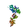
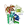
- PDB-2p8z: Fitted structure of ADPR-eEF2 in the 80S:ADPR-eEF2:GDPNP:sordarin... -
+ Open data

- Basic information
| Entry | Database: PDB / ID: 2p8z | ||||||
|---|---|---|---|---|---|---|---|
| Title | Fitted structure of ADPR-eEF2 in the 80S:ADPR-eEF2:GDPNP:sordarin cryo-EM reconstruction | ||||||
 Components Components |
| ||||||
 Keywords Keywords | TRANSLATION / elongation / translocation / GTPase / 80S ribosome | ||||||
| Function / homology |  Function and homology information Function and homology informationPeptide chain elongation / Synthesis of diphthamide-EEF2 / positive regulation of translational elongation / Protein methylation / translational elongation / translation elongation factor activity / Neutrophil degranulation / maintenance of translational fidelity / Hydrolases; Acting on acid anhydrides; Acting on GTP to facilitate cellular and subcellular movement / ribosome binding ...Peptide chain elongation / Synthesis of diphthamide-EEF2 / positive regulation of translational elongation / Protein methylation / translational elongation / translation elongation factor activity / Neutrophil degranulation / maintenance of translational fidelity / Hydrolases; Acting on acid anhydrides; Acting on GTP to facilitate cellular and subcellular movement / ribosome binding / protein-folding chaperone binding / rRNA binding / ribonucleoprotein complex / GTPase activity / GTP binding / identical protein binding / cytosol / cytoplasm Similarity search - Function | ||||||
| Biological species |    Thermus thermophilus (bacteria) Thermus thermophilus (bacteria) | ||||||
| Method | ELECTRON MICROSCOPY / single particle reconstruction / cryo EM / Resolution: 8.9 Å | ||||||
 Authors Authors | Taylor, D.J. / Nilsson, J. / Merrill, A.R. / Andersen, G.R. / Nissen, P. / Frank, J. | ||||||
 Citation Citation | Journal: EMBO J / Year: 2007 Title: Structures of modified eEF2 80S ribosome complexes reveal the role of GTP hydrolysis in translocation. Authors: Derek J Taylor / Jakob Nilsson / A Rod Merrill / Gregers Rom Andersen / Poul Nissen / Joachim Frank /  Abstract: On the basis of kinetic data on ribosome protein synthesis, the mechanical energy for translocation of the mRNA-tRNA complex is thought to be provided by GTP hydrolysis of an elongation factor (eEF2 ...On the basis of kinetic data on ribosome protein synthesis, the mechanical energy for translocation of the mRNA-tRNA complex is thought to be provided by GTP hydrolysis of an elongation factor (eEF2 in eukaryotes, EF-G in bacteria). We have obtained cryo-EM reconstructions of eukaryotic ribosomes complexed with ADP-ribosylated eEF2 (ADPR-eEF2), before and after GTP hydrolysis, providing a structural basis for analyzing the GTPase-coupled mechanism of translocation. Using the ADP-ribosyl group as a distinct marker, we observe conformational changes of ADPR-eEF2 that are due strictly to GTP hydrolysis. These movements are likely representative of native eEF2 motions in a physiological context and are sufficient to uncouple the mRNA-tRNA complex from two universally conserved bases in the ribosomal decoding center (A1492 and A1493 in Escherichia coli) during translocation. Interpretation of these data provides a detailed two-step model of translocation that begins with the eEF2/EF-G binding-induced ratcheting motion of the small ribosomal subunit. GTP hydrolysis then uncouples the mRNA-tRNA complex from the decoding center so translocation of the mRNA-tRNA moiety may be completed by a head rotation of the small subunit. | ||||||
| History |
|
- Structure visualization
| Movie |
Movie viewer |
|---|---|
| Structure viewer | Molecule: MolmilJmol/JSmol |
- Downloads & links
-Download
| PDBx/mmCIF format | 2p8z.cif.gz | 158.9 KB | Display | PDBx/mmCIF format |
|---|---|---|---|---|
| PDB format | pdb2p8z.ent.gz | 125.5 KB | Display | PDB format |
| PDBx/mmJSON format | 2p8z.json.gz | Tree view | PDBx/mmJSON format | |
| Others |  Other downloads Other downloads |
-Validation report
| Summary document | 2p8z_validation.pdf.gz | 962.5 KB | Display | wwPDB validaton report |
|---|---|---|---|---|
| Full document | 2p8z_full_validation.pdf.gz | 1.1 MB | Display | |
| Data in XML | 2p8z_validation.xml.gz | 41 KB | Display | |
| Data in CIF | 2p8z_validation.cif.gz | 57.3 KB | Display | |
| Arichive directory | https://data.pdbj.org/pub/pdb/validation_reports/p8/2p8zftp://data.pdbj.org/pub/pdb/validation_reports/p8/2p8z | HTTPS FTP |
-Related structure data
| Related structure data | 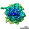 1345MC 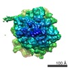 1342C 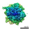 1343C 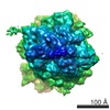 1344C  2p8wC  2p8xC 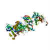 2p8yC C: citing same article ( M: map data used to model this data |
|---|---|
| Similar structure data |


-Links
 PDBj
PDBj


- Assembly
| Deposited unit | 
|
|---|---|
| 1 |
|
-Components
| #1: Protein | Mass: 93549.320 Da / Num. of mol.: 1 / Source method: isolated from a natural source / Source: (natural) |
|---|---|
| #2: Protein/peptide | Mass: 3829.122 Da / Num. of mol.: 1 / Fragment: SWITCH 1 LOOP / Source method: isolated from a natural source / Source: (natural) Thermus thermophilus (bacteria) / References: UniProt: P60339 |
| #3: Chemical | ChemComp-APR /   Mass: 559.316 Da / Num. of mol.: 1 / Source method: obtained synthetically / Formula: C15H23N5O14P2 Mass: 559.316 Da / Num. of mol.: 1 / Source method: obtained synthetically / Formula: C15H23N5O14P2 |
| #4: Chemical | ChemComp-SO1 / [  Mass: 494.618 Da / Num. of mol.: 1 / Source method: obtained synthetically / Formula: C27H42O8 Mass: 494.618 Da / Num. of mol.: 1 / Source method: obtained synthetically / Formula: C27H42O8 |
| #5: Chemical | ChemComp-GNP /   Mass: 522.196 Da / Num. of mol.: 1 / Source method: obtained synthetically / Formula: C10H17N6O13P3 Mass: 522.196 Da / Num. of mol.: 1 / Source method: obtained synthetically / Formula: C10H17N6O13P3Comment: GppNHp, GMPPNP, energy-carrying molecule analogue*YM |
-Experimental details
-Experiment
| Experiment | Method: ELECTRON MICROSCOPY |
|---|---|
| EM experiment | Aggregation state: PARTICLE / 3D reconstruction method: single particle reconstruction |
- Sample preparation
| Component | Name: 80S ribosome / Type: RIBOSOME |
|---|---|
| Buffer solution | pH: 7.5 |
| Specimen | Conc.: 0.096 mg/ml / Embedding applied: NO / Shadowing applied: NO / Staining applied: NO / Vitrification applied: YES |
| Specimen support | Details: Quantifoil holey-Carbon film grids |
| Vitrification | Instrument: FEI VITROBOT MARK I / Cryogen name: ETHANE / Details: rapid-freezing in liquid ethane |
- Electron microscopy imaging
| Experimental equipment |  Model: Tecnai F30 / Image courtesy: FEI Company |
|---|---|
| Microscopy | Model: FEI TECNAI F30 / Date: Jul 7, 2005 |
| Electron gun | Electron source:  FIELD EMISSION GUN / Accelerating voltage: 300 kV / Illumination mode: FLOOD BEAM FIELD EMISSION GUN / Accelerating voltage: 300 kV / Illumination mode: FLOOD BEAM |
| Electron lens | Mode: BRIGHT FIELD / Nominal magnification: 39000 X / Calibrated magnification: 37642 X / Nominal defocus max: 4000 nm / Nominal defocus min: 1400 nm / Cs: 2.26 mm |
| Specimen holder | Temperature: 93 K / Tilt angle max: 0 ° / Tilt angle min: 0 ° |
| Image recording | Electron dose: 25 e/Å2 / Film or detector model: KODAK SO-163 FILM |
| Radiation | Protocol: SINGLE WAVELENGTH / Monochromatic (M) / Laue (L): M |
| Radiation wavelength | Relative weight: 1 |
- Processing
| EM software |
| ||||||||||||
|---|---|---|---|---|---|---|---|---|---|---|---|---|---|
| CTF correction | Details: CTF correction of 3D-maps by Wiener filtration | ||||||||||||
| Symmetry | Point symmetry: C1 (asymmetric) | ||||||||||||
| 3D reconstruction | Method: 3D projection matching, conjugate gradients with regularization Resolution: 8.9 Å / Num. of particles: 102689 / Nominal pixel size: 1.86 Å / Actual pixel size: 1.86 Å / Magnification calibration: TMV / Details: SPIDER package / Symmetry type: POINT | ||||||||||||
| Atomic model building | B value: 15 / Protocol: RIGID BODY FIT / Space: REAL / Target criteria: Maximization of correlation-coefficient Details: METHOD--Real-space refinement using rigid bodies REFINEMENT PROTOCOL--TNT implementation of RSRef | ||||||||||||
| Atomic model building |
| ||||||||||||
| Refinement step | Cycle: LAST
|