 Movie
Movie Controller

[English] 日本語
 Yorodumi
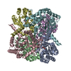
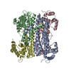
- PDB-2f3r: Crystal Structure Of E.coli Guanylate Kinase In Complex With Ap5G -
+ Open data

- Basic information
| Entry | Database: PDB / ID: 2f3r | ||||||
|---|---|---|---|---|---|---|---|
| Title | Crystal Structure Of E.coli Guanylate Kinase In Complex With Ap5G | ||||||
 Components Components | Guanylate kinase | ||||||
 Keywords Keywords | TRANSFERASE / GMP KINASE / GUANYLATE KINASE / NUCLEOTIDE ANALOGUE | ||||||
| Function / homology |  Function and homology information Function and homology informationguanylate kinase / GMP kinase activity / ATP binding / identical protein binding / cytosol Similarity search - Function | ||||||
| Biological species |  | ||||||
| Method |  X-RAY DIFFRACTION / SYNCHROTRON / FOURIER SYNTHESIS / Resolution: 2.5 Å X-RAY DIFFRACTION / SYNCHROTRON / FOURIER SYNTHESIS / Resolution: 2.5 Å | ||||||
 Authors Authors | Hible, G. / Cherfils, J. | ||||||
 Citation Citation | Journal: Biochimie / Year: 2006 Title: Crystal structures of GMP kinase in complex with ganciclovir monophosphate and Ap5G. Authors: Hible, G. / Daalova, P. / Gilles, A.M. / Cherfils, J. | ||||||
| History |
|
- Structure visualization
| Structure viewer | Molecule: MolmilJmol/JSmol |
|---|
- Downloads & links
-Download
| PDBx/mmCIF format | 2f3r.cif.gz | 95.2 KB | Display | PDBx/mmCIF format |
|---|---|---|---|---|
| PDB format | pdb2f3r.ent.gz | 73.1 KB | Display | PDB format |
| PDBx/mmJSON format | 2f3r.json.gz | Tree view | PDBx/mmJSON format | |
| Others |  Other downloads Other downloads |
-Validation report
| Arichive directory | https://data.pdbj.org/pub/pdb/validation_reports/f3/2f3rftp://data.pdbj.org/pub/pdb/validation_reports/f3/2f3r | HTTPS FTP |
|---|
-Related structure data
| Related structure data | 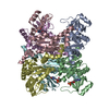 2f3tC  2an9S C: citing same article ( S: Starting model for refinement |
|---|---|
| Similar structure data |


-Links
 PDBj
PDBj- Assembly
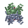
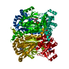
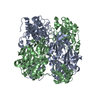
| Deposited unit | 
| ||||||||
|---|---|---|---|---|---|---|---|---|---|
| 1 | 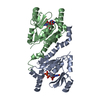
| ||||||||
| Unit cell |
| ||||||||
| Details | THIS ENTRY CONTAINS THE CRYSTALLOGRAPHIC ASYMMETRIC UNIT WHICH CONSISTS OF A DIMER (CHAINS A and B). The biological assembly is a hexamer generated from the dimer in the asymmetric unit by the operations: -y, x-y, z and -x+y, -x, z. |
-Components
| #1: Protein | Mass: 23625.725 Da / Num. of mol.: 2 Source method: isolated from a genetically manipulated source Source: (gene. exp.) #2: Chemical |   Mass: 932.366 Da / Num. of mol.: 2 / Source method: obtained synthetically / Formula: C20H29N10O23P5 Mass: 932.366 Da / Num. of mol.: 2 / Source method: obtained synthetically / Formula: C20H29N10O23P5#3: Water | ChemComp-HOH / |  Mass: 18.015 Da / Num. of mol.: 26 / Source method: isolated from a natural source / Formula: H2O Mass: 18.015 Da / Num. of mol.: 26 / Source method: isolated from a natural source / Formula: H2O |
|---|
-Experimental details
-Experiment
| Experiment | Method: X-RAY DIFFRACTION / Number of used crystals: 1 |
|---|
- Sample preparation
| Crystal | Density Matthews: 2.08 Å3/Da / Density % sol: 41 % |
|---|---|
| Crystal grow | Temperature: 292 K / Method: vapor diffusion, sitting drop / pH: 6.5 Details: 30% PEG 4000, 100mM MES, pH 6.5, VAPOR DIFFUSION, SITTING DROP, temperature 292K |
-Data collection
| Diffraction | Mean temperature: 100 K |
|---|---|
| Diffraction source | Source: SYNCHROTRON / Site: ESRF  / Beamline: ID14-1 / Wavelength: 0.934 Å / Beamline: ID14-1 / Wavelength: 0.934 Å |
| Detector | Type: ADSC QUANTUM 4 / Detector: CCD / Date: Sep 18, 2005 |
| Radiation | Protocol: SINGLE WAVELENGTH / Monochromatic (M) / Laue (L): M / Scattering type: x-ray |
| Radiation wavelength | Wavelength: 0.934 Å / Relative weight: 1 |
| Reflection | Resolution: 2.5→28 Å / Num. all: 13344 / Num. obs: 13344 / % possible obs: 99.4 % / Observed criterion σ(F): 0 / Observed criterion σ(I): 0 / Redundancy: 11.4 % / Biso Wilson estimate: 41.3 Å2 / Rmerge(I) obs: 0.064 / Net I/σ(I): 27.2 |
| Reflection shell | Resolution: 2.5→2.66 Å / Redundancy: 11.5 % / Rmerge(I) obs: 0.399 / Mean I/σ(I) obs: 7 / Num. unique all: 2192 / % possible all: 100 |
- Processing
| Software |
| ||||||||||||||||||||||||||||||||||||
|---|---|---|---|---|---|---|---|---|---|---|---|---|---|---|---|---|---|---|---|---|---|---|---|---|---|---|---|---|---|---|---|---|---|---|---|---|---|
| Refinement | Method to determine structure: FOURIER SYNTHESIS Starting model: PDB ENTRY 2AN9 Resolution: 2.5→27.96 Å / Rfactor Rfree error: 0.01 / Data cutoff high absF: 1728695.83 / Data cutoff low absF: 0 / Isotropic thermal model: RESTRAINED / Cross valid method: THROUGHOUT / σ(F): 0 / Stereochemistry target values: Engh & Huber
| ||||||||||||||||||||||||||||||||||||
| Solvent computation | Solvent model: FLAT MODEL / Bsol: 31.4334 Å2 / ksol: 0.312595 e/Å3 | ||||||||||||||||||||||||||||||||||||
| Displacement parameters | Biso mean: 52.8 Å2
| ||||||||||||||||||||||||||||||||||||
| Refine analyze |
| ||||||||||||||||||||||||||||||||||||
| Refinement step | Cycle: LAST / Resolution: 2.5→27.96 Å
| ||||||||||||||||||||||||||||||||||||
| Refine LS restraints |
| ||||||||||||||||||||||||||||||||||||
| LS refinement shell | Resolution: 2.5→2.66 Å / Rfactor Rfree error: 0.032 / Total num. of bins used: 6
| ||||||||||||||||||||||||||||||||||||
| Xplor file |
|
