SHEET DETERMINATION METHOD: DSSP THE SHEETS PRESENTED AS "AB" IN EACH CHAIN ON SHEET RECORDS BELOW ... SHEET DETERMINATION METHOD: DSSP THE SHEETS PRESENTED AS "AB" IN EACH CHAIN ON SHEET RECORDS BELOW IS ACTUALLY AN 8-STRANDED BARREL THIS IS REPRESENTED BY A 9-STRANDED SHEET IN WHICH THE FIRST AND LAST STRANDS ARE IDENTICAL. THE SHEETS PRESENTED AS "BB" IN EACH CHAIN ON SHEET RECORDS BELOW IS ACTUALLY AN 8-STRANDED BARREL THIS IS REPRESENTED BY A 9-STRANDED SHEET IN WHICH THE FIRST AND LAST STRANDS ARE IDENTICAL.
Mass: 18.015 Da / Num. of mol.: 650 / Source method: isolated from a natural source / Formula: H2O
-
Details
Sequence details
ACCORDING TO THE AUTHORS THE CONFLICTS IN SEQADV ARE ALL VARIANTS
-
Experimental details
-
Experiment
Experiment
Method: X-RAY DIFFRACTION / Number of used crystals: 1
-
Sample preparation
Crystal
Density Matthews: 2.51 Å3/Da / Density % sol: 47.5 %
Crystal grow
pH: 6.5 Details: 0.2 M AMMONIUM SULFATE, 0.1 M SODIUM CACODYLATE PH 6.5, 30 % PEG 8000) AND 0.25 MICROLITER OF 40 % V/V GAMMA-BUTYROLACTONE TO A 1 PLUS MICROLITER DROP
Monochromator: SI(111) / Protocol: SINGLE WAVELENGTH / Monochromatic (M) / Laue (L): M / Scattering type: x-ray
Radiation wavelength
Wavelength: 1.28202 Å / Relative weight: 1
Reflection
Resolution: 2.25→20 Å / Num. obs: 65956 / % possible obs: 100 % / Observed criterion σ(I): 2 / Redundancy: 25.4 % / Rmerge(I) obs: 0.11 / Net I/σ(I): 7.7
Reflection shell
Resolution: 2.25→2.33 Å / Redundancy: 18.6 % / Rmerge(I) obs: 0.48 / Mean I/σ(I) obs: 7.6 / % possible all: 100
-
Processing
Software
Name
Version
Classification
REFMAC
5.2.0005
refinement
DENZO
datareduction
SCALEPACK
datascaling
SOLVE
phasing
Refinement
Method to determine structure: SAD / Resolution: 2.25→20 Å / Cor.coef. Fo:Fc: 0.953 / Cor.coef. Fo:Fc free: 0.912 / SU B: 10.388 / SU ML: 0.139 / Cross valid method: THROUGHOUT / ESU R: 0.238 / ESU R Free: 0.194 / Stereochemistry target values: MAXIMUM LIKELIHOOD Details: HYDROGENS HAVE BEEN ADDED IN THE RIDING POSITIONS. RESIDUES 31 TO 40 ARE DISORDERED
Rfactor
Num. reflection
% reflection
Selection details
Rfree
0.22
659
1 %
RANDOM
Rwork
0.169
-
-
-
obs
0.169
65296
99.9 %
-
Solvent computation
Ion probe radii: 0.8 Å / Shrinkage radii: 0.8 Å / VDW probe radii: 1.2 Å / Solvent model: BABINET MODEL WITH MASK
 Movie
Movie Controller

 Yorodumi
 Open data
 Basic information
 Components
 Keywords
 Function and homology information
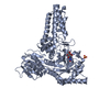
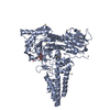
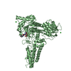
 CLOSTRIDIUM PERFRINGENS (bacteria)
 X-RAY DIFFRACTION /
  Authors
 Citation
 Structure visualization
 Downloads & links
 Other downloads


 PDBj
PDBj


 Assembly


 Mass: 96.063 Da / Num. of mol.: 1 / Source method: obtained synthetically / Formula: SO4
 Mass: 35.453 Da / Num. of mol.: 1 / Source method: obtained synthetically / Formula: Cl
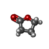
 Mass: 86.089 Da / Num. of mol.: 2 / Source method: obtained synthetically / Formula: C4H6O2
 Mass: 92.094 Da / Num. of mol.: 2 / Source method: obtained synthetically / Formula: C3H8O3
 Mass: 65.409 Da / Num. of mol.: 11 / Source method: obtained synthetically / Formula: Zn
 Sample preparation
 / Beamline: BM14 / Wavelength: 1.28202
  Processing