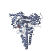
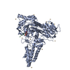
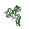
Entry Database : PDB / ID : 2vurTitle Chemical dissection of the link between Streptozotocin, O-GlcNAc and pancreatic cell death O-GLCNACASE NAGJ Keywords / / / Function / homology Function Domain/homology Component
/ / / / / / / / / / / / / / / / / / / / / / / / / / / / / / / / / / / / / / / / / / / / / / / Biological species CLOSTRIDIUM PERFRINGENS (bacteria)Method / / / Resolution : 2.2 Å Authors Pathak, S. / Dorfmueller, H.C. / Borodkin, V.S. / van Aalten, D.M.F. Journal : Chem.Biol. / Year : 2008Title : Chemical Dissection of the Link between Streptozotocin, O-Glcnac, and Pancreatic Cell Death.Authors : Pathak, S. / Dorfmueller, H.C. / Borodkin, V.S. / Van Aalten, D.M.F. History Deposition May 29, 2008 Deposition site / Processing site Revision 1.0 Feb 10, 2009 Provider / Type Revision 1.1 Jul 13, 2011 Group / Version format complianceRevision 1.2 Jul 29, 2020 Group / Derived calculations / OtherCategory chem_comp / pdbx_database_status ... chem_comp / pdbx_database_status / struct_site / struct_site_gen Item / _pdbx_database_status.status_code_sfDescription / Provider / Type Revision 1.3 May 8, 2024 Group / Database references / Structure summaryCategory chem_comp / chem_comp_atom ... chem_comp / chem_comp_atom / chem_comp_bond / database_2 Item / _database_2.pdbx_DOI / _database_2.pdbx_database_accession
Show all Show less Remark 700 SHEET DETERMINATION METHOD: DSSP THE SHEETS PRESENTED AS "AC" IN EACH CHAIN ON SHEET RECORDS BELOW ... SHEET DETERMINATION METHOD: DSSP THE SHEETS PRESENTED AS "AC" IN EACH CHAIN ON SHEET RECORDS BELOW IS ACTUALLY AN 8-STRANDED BARREL THIS IS REPRESENTED BY A 9-STRANDED SHEET IN WHICH THE FIRST AND LAST STRANDS ARE IDENTICAL. THE SHEETS PRESENTED AS "BC" IN EACH CHAIN ON SHEET RECORDS BELOW IS ACTUALLY AN 8-STRANDED BARREL THIS IS REPRESENTED BY A 9-STRANDED SHEET IN WHICH THE FIRST AND LAST STRANDS ARE IDENTICAL.
 Movie
Movie Controller

 Yorodumi
 Open data
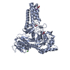
 Basic information
 Components
 Keywords
 Function and homology information
 CLOSTRIDIUM PERFRINGENS (bacteria)
 X-RAY DIFFRACTION /
  Authors
 Citation
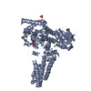
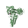
 Structure visualization
 Downloads & links
 Other downloads


 PDBj
PDBj


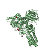
 Assembly
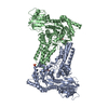
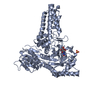
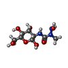
 Type: D-saccharide / Mass: 267.236 Da / Num. of mol.: 2 / Source method: obtained synthetically / Formula: C8H17N3O7
 Mass: 96.063 Da / Num. of mol.: 1 / Source method: obtained synthetically / Formula: SO4
 Mass: 18.015 Da / Num. of mol.: 528 / Source method: isolated from a natural source / Formula: H2O
 Sample preparation
 / Beamline: ID14-4 / Wavelength: 0.931
  Processing