 Movie
Movie Controller


+ Open data

- Basic information
| Entry | Database: PDB / ID: 2c1l | ||||||
|---|---|---|---|---|---|---|---|
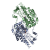
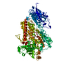
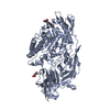
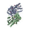
| Title | Structure of the BfiI restriction endonuclease | ||||||
 Components Components | RESTRICTION ENDONUCLEASE | ||||||
 Keywords Keywords | HYDROLASE / BFII / RESTRICTION ENDONUCLEASE / DOMAIN FUSION | ||||||
| Function / homology |  Function and homology information Function and homology information | ||||||
| Biological species |  BACILLUS FIRMUS (bacteria) BACILLUS FIRMUS (bacteria) | ||||||
| Method |  X-RAY DIFFRACTION / SYNCHROTRON / MIRAS / Resolution: 1.9 Å X-RAY DIFFRACTION / SYNCHROTRON / MIRAS / Resolution: 1.9 Å | ||||||
 Authors Authors | Grazulis, S. / Manakova, E. / Roessle, M. / Bochtler, M. / Tamulaitiene, G. / Huber, R. / Siksnys, V. | ||||||
 Citation Citation | Journal: Proc.Natl.Acad.Sci.USA / Year: 2005 Title: Structure of the Metal-Independent Restriction Enzyme Bfii Reveals Fusion of a Specific DNA-Binding Domain with a Nonspecific Nuclease. Authors: Grazulis, S. / Manakova, E. / Roessle, M. / Bochtler, M. / Tamulaitiene, G. / Huber, R. / Siksnys, V. | ||||||
| History |
| ||||||
| Remark 700 | SHEET THE SHEET STRUCTURE OF THIS MOLECULE IS BIFURCATED. IN ORDER TO REPRESENT THIS FEATURE IN ... SHEET THE SHEET STRUCTURE OF THIS MOLECULE IS BIFURCATED. IN ORDER TO REPRESENT THIS FEATURE IN THE SHEET RECORDS BELOW, TWO SHEETS ARE DEFINED. |
- Structure visualization
| Structure viewer | Molecule: MolmilJmol/JSmol |
|---|
- Downloads & links
-Download
| PDBx/mmCIF format | 2c1l.cif.gz | 165.9 KB | Display | PDBx/mmCIF format |
|---|---|---|---|---|
| PDB format | pdb2c1l.ent.gz | 134 KB | Display | PDB format |
| PDBx/mmJSON format | 2c1l.json.gz | Tree view | PDBx/mmJSON format | |
| Others |  Other downloads Other downloads |
-Validation report
| Arichive directory | https://data.pdbj.org/pub/pdb/validation_reports/c1/2c1lftp://data.pdbj.org/pub/pdb/validation_reports/c1/2c1l | HTTPS FTP |
|---|
-Related structure data
| Similar structure data |
|---|


-Links
 PDBj
PDBj


- Assembly
| Deposited unit | 
| ||||||||
|---|---|---|---|---|---|---|---|---|---|
| 1 |
| ||||||||
| Unit cell |
|
-Components
-Protein , 1 types, 2 molecules AB
| #1: Protein | Mass: 40041.566 Da / Num. of mol.: 2 Source method: isolated from a genetically manipulated source Source: (gene. exp.) BACILLUS FIRMUS (bacteria) / Strain: S8120 / Plasmid: PET21B-BFIIR6, 5 / Production host: References: UniProt: Q9F4C9, type II site-specific deoxyribonuclease |
|---|
-Non-polymers , 8 types, 494 molecules 


| #2: Chemical | ChemComp-GOL /  Mass: 92.094 Da / Num. of mol.: 4 / Source method: obtained synthetically / Formula: C3H8O3 Mass: 92.094 Da / Num. of mol.: 4 / Source method: obtained synthetically / Formula: C3H8O3#3: Chemical | ChemComp-TRS / |  Mass: 122.143 Da / Num. of mol.: 1 / Source method: obtained synthetically / Formula: C4H12NO3 / Comment: pH buffer*YM Mass: 122.143 Da / Num. of mol.: 1 / Source method: obtained synthetically / Formula: C4H12NO3 / Comment: pH buffer*YM#4: Chemical |  Mass: 150.087 Da / Num. of mol.: 2 / Source method: obtained synthetically / Formula: C4H6O6 Mass: 150.087 Da / Num. of mol.: 2 / Source method: obtained synthetically / Formula: C4H6O6#5: Chemical |  Mass: 150.087 Da / Num. of mol.: 2 / Source method: obtained synthetically / Formula: C4H6O6 Mass: 150.087 Da / Num. of mol.: 2 / Source method: obtained synthetically / Formula: C4H6O6#6: Chemical | ChemComp-TAR / |  Mass: 150.087 Da / Num. of mol.: 1 / Source method: obtained synthetically / Formula: C4H6O6 Mass: 150.087 Da / Num. of mol.: 1 / Source method: obtained synthetically / Formula: C4H6O6#7: Chemical |  Mass: 61.017 Da / Num. of mol.: 2 / Source method: obtained synthetically / Formula: CHO3 / Comment: pH buffer*YM Mass: 61.017 Da / Num. of mol.: 2 / Source method: obtained synthetically / Formula: CHO3 / Comment: pH buffer*YM#8: Chemical |  Mass: 195.237 Da / Num. of mol.: 2 / Source method: obtained synthetically / Formula: C6H13NO4S / Comment: pH buffer*YM Mass: 195.237 Da / Num. of mol.: 2 / Source method: obtained synthetically / Formula: C6H13NO4S / Comment: pH buffer*YM#9: Water | ChemComp-HOH / | Mass: 18.015 Da / Num. of mol.: 480 / Source method: isolated from a natural source / Formula: H2O |
|---|
-Experimental details
-Experiment
| Experiment | Method: X-RAY DIFFRACTION / Number of used crystals: 1 |
|---|
- Sample preparation
| Crystal | Density Matthews: 2.51 Å3/Da / Density % sol: 50.66 % |
|---|---|
| Crystal grow | pH: 6.5 / Details: MES 0.1M PH 6.5 K/NA TARTRATE 1.1M GLYCEROL 25% |
-Data collection
| Diffraction | Mean temperature: 100 K |
|---|---|
| Diffraction source | Source: SYNCHROTRON / Site: ESRF  / Beamline: ID13 / Wavelength: 0.9755 / Beamline: ID13 / Wavelength: 0.9755 |
| Detector | Type: MARRESEARCH / Detector: CCD / Date: Sep 9, 2001 / Details: MIRRORS |
| Radiation | Monochromator: SINGLE CRYSTAL / Protocol: SINGLE WAVELENGTH / Monochromatic (M) / Laue (L): M / Scattering type: x-ray |
| Radiation wavelength | Wavelength: 0.9755 Å / Relative weight: 1 |
| Reflection | Resolution: 1.9→28 Å / Num. obs: 70374 / % possible obs: 99.9 % / Observed criterion σ(I): 0 / Redundancy: 3.8 % / Biso Wilson estimate: 15.1 Å2 / Rmerge(I) obs: 0.05 / Net I/σ(I): 10.4 |
| Reflection shell | Resolution: 1.9→2 Å / Redundancy: 3.8 % / Rmerge(I) obs: 0.26 / Mean I/σ(I) obs: 2.7 / % possible all: 100 |
- Processing
| Software |
| ||||||||||||||||||||||||||||||||||||||||||||||||||||||||||||||||||||||||||||||||
|---|---|---|---|---|---|---|---|---|---|---|---|---|---|---|---|---|---|---|---|---|---|---|---|---|---|---|---|---|---|---|---|---|---|---|---|---|---|---|---|---|---|---|---|---|---|---|---|---|---|---|---|---|---|---|---|---|---|---|---|---|---|---|---|---|---|---|---|---|---|---|---|---|---|---|---|---|---|---|---|---|---|
| Refinement | Method to determine structure: MIRAS / Resolution: 1.9→28.01 Å / Rfactor Rfree error: 0.003 / Data cutoff high absF: 31299524.91 / Isotropic thermal model: RESTRAINED / Cross valid method: THROUGHOUT / σ(F): 0 / Stereochemistry target values: RESIDUAL Details: RESOLUTION-DEPENDENT WEIGHTING SCHEME USED. BULK SOLVENT MODEL USED
| ||||||||||||||||||||||||||||||||||||||||||||||||||||||||||||||||||||||||||||||||
| Solvent computation | Solvent model: FLAT MODEL / Bsol: 69.1296 Å2 / ksol: 0.382458 e/Å3 | ||||||||||||||||||||||||||||||||||||||||||||||||||||||||||||||||||||||||||||||||
| Displacement parameters | Biso mean: 27.2 Å2
| ||||||||||||||||||||||||||||||||||||||||||||||||||||||||||||||||||||||||||||||||
| Refine analyze |
| ||||||||||||||||||||||||||||||||||||||||||||||||||||||||||||||||||||||||||||||||
| Refinement step | Cycle: LAST / Resolution: 1.9→28.01 Å
| ||||||||||||||||||||||||||||||||||||||||||||||||||||||||||||||||||||||||||||||||
| Refine LS restraints |
| ||||||||||||||||||||||||||||||||||||||||||||||||||||||||||||||||||||||||||||||||
| LS refinement shell | Resolution: 1.9→2.02 Å / Rfactor Rfree error: 0.008 / Total num. of bins used: 6
| ||||||||||||||||||||||||||||||||||||||||||||||||||||||||||||||||||||||||||||||||
| Xplor file |
|
