National Institutes of Health/National Institute of General Medical Sciences (NIH/NIGMS)
GM114611
United States
National Institutes of Health/National Institute of General Medical Sciences (NIH/NIGMS)
GM105385
United States
Citation
Journal: Science / Year: 2023 Title: The mechanism of the phage-encoded protein antibiotic from ΦX174. Authors: Anna K Orta / Nadia Riera / Yancheng E Li / Shiho Tanaka / Hyun Gi Yun / Lada Klaic / William M Clemons / Abstract: The historically important phage ΦX174 kills its host bacteria by encoding a 91-residue protein antibiotic called protein E. Using single-particle electron cryo-microscopy, we demonstrate that ...The historically important phage ΦX174 kills its host bacteria by encoding a 91-residue protein antibiotic called protein E. Using single-particle electron cryo-microscopy, we demonstrate that protein E bridges two bacterial proteins to form the transmembrane YES complex [MraY, protein E, sensitivity to lysis D (SlyD)]. Protein E inhibits peptidoglycan biosynthesis by obstructing the MraY active site leading to loss of lipid I production. We experimentally validate this result for two different viral species, providing a clear model for bacterial lysis and unifying previous experimental data. Additionally, we characterize the MraY structure-revealing features of this essential enzyme-and the structure of the chaperone SlyD bound to a protein. Our structures provide insights into the mechanism of phage-mediated lysis and for structure-based design of phage therapeutics.
Protein or peptide: Phospho-N-acetylmuramoyl-pentapeptide-transferase
Complex: SlyD
Protein or peptide: Peptidyl-prolyl cis-trans isomerase
-
Supramolecule #1: YES complex
Supramolecule
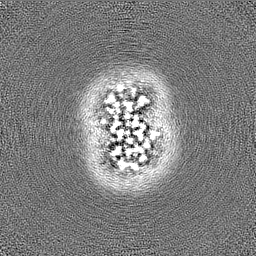
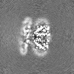
Name: YES complex / type: complex / ID: 1 / Parent: 0 / Macromolecule list: all Details: Hexameric complex of E. coli MraY dimer bound to two molecules of Protein E (phix174), stabilized by E. coli SlyD
Molecular weight
Theoretical: 140.32 KDa
-
Supramolecule #2: Lysis Protein E
Supramolecule
Name: Lysis Protein E / type: complex / ID: 2 / Parent: 1 / Macromolecule list: #2 / Details: Protein E from PhiX174 phage
Details: Supplemented with 2mM E. coli lipid extract
Grid
Model: Quantifoil R1.2/1.3 / Material: COPPER / Mesh: 300 / Support film - Material: CARBON / Support film - topology: HOLEY
Vitrification
Cryogen name: ETHANE / Chamber humidity: 100 % / Chamber temperature: 277.15 K / Instrument: FEI VITROBOT MARK IV
-
Electron microscopy
Microscope
FEI TITAN KRIOS
Image recording
Film or detector model: GATAN K3 (6k x 4k) / Number grids imaged: 1 / Number real images: 10798 / Average exposure time: 2.0 sec. / Average electron dose: 60.0 e/Å2
Electron beam
Acceleration voltage: 300 kV / Electron source: FIELD EMISSION GUN
In the structure databanks used in Yorodumi, some data are registered as the other names, "COVID-19 virus" and "2019-nCoV". Here are the details of the virus and the list of structure data.
Jan 31, 2019. EMDB accession codes are about to change! (news from PDBe EMDB page)
EMDB accession codes are about to change! (news from PDBe EMDB page)
The allocation of 4 digits for EMDB accession codes will soon come to an end. Whilst these codes will remain in use, new EMDB accession codes will include an additional digit and will expand incrementally as the available range of codes is exhausted. The current 4-digit format prefixed with “EMD-” (i.e. EMD-XXXX) will advance to a 5-digit format (i.e. EMD-XXXXX), and so on. It is currently estimated that the 4-digit codes will be depleted around Spring 2019, at which point the 5-digit format will come into force.
The EM Navigator/Yorodumi systems omit the EMD- prefix.
Related info.:Q: What is EMD? / ID/Accession-code notation in Yorodumi/EM Navigator
Yorodumi is a browser for structure data from EMDB, PDB, SASBDB, etc.
This page is also the successor to EM Navigator detail page, and also detail information page/front-end page for Omokage search.
The word "yorodu" (or yorozu) is an old Japanese word meaning "ten thousand". "mi" (miru) is to see.
Related info.:EMDB / PDB / SASBDB / Comparison of 3 databanks / Yorodumi Search / Aug 31, 2016. New EM Navigator & Yorodumi / Yorodumi Papers / Jmol/JSmol / Function and homology information / Changes in new EM Navigator and Yorodumi
 Movie
Movie Controller


 Open data
 Basic information
 Map data
 Sample
 Keywords
 Function and homology information
 Escherichia phage phiX174 (virus) /
 Authors
 United States, 2 items
  Citation
 Structure visualization
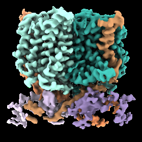
 Downloads & links
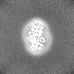
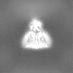
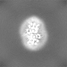
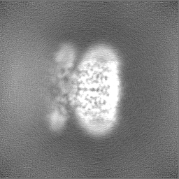
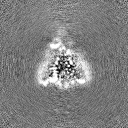
 emd_29642.png
 http://ftp.pdbj.org/pub/emdb/structures/EMD-29642


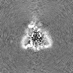
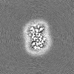
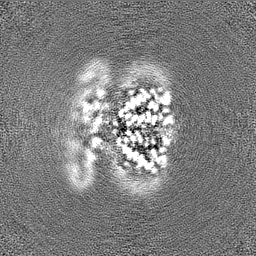
 Z
Z Y
Y X
X


 Sample components
 Processing
 Electron microscopy
 FIELD EMISSION GUN