[English] 日本語
 Yorodumi
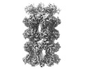
- EMDB-28965: Glutamine synthetase from Pseudomonas aeruginosa, filament double... -
+ Open data
- Basic information
| Entry |  | |||||||||
|---|---|---|---|---|---|---|---|---|---|---|
| Title | Glutamine synthetase from Pseudomonas aeruginosa, filament double-unit in compressed conformation | |||||||||
 Map data Map data | ||||||||||
 Sample Sample |
| |||||||||
 Keywords Keywords | glutamine biosynthetic process nitrogen utilization / Structural Genomics / Seattle Structural Genomics Center for Infectious Disease / SSGCID / LIGASE | |||||||||
| Function / homology |  Function and homology information Function and homology informationnitrogen utilization / glutamine synthetase / glutamine biosynthetic process / glutamine synthetase activity / ATP binding / membrane / metal ion binding / cytoplasm Similarity search - Function | |||||||||
| Biological species |   Pseudomonas aeruginosa PAO1 (bacteria) Pseudomonas aeruginosa PAO1 (bacteria) | |||||||||
| Method | single particle reconstruction / cryo EM / Resolution: 2.8 Å | |||||||||
 Authors Authors | Phan IQ / Staker B / Shek R / Moser TH / Evans JE / van Voorhis WC / Myler PJ / Seattle Structural Genomics Center for Infectious Disease (SSGCID) | |||||||||
| Funding support |  United States, 1 items United States, 1 items
| |||||||||
 Citation Citation |  Journal: To Be Published Journal: To Be PublishedTitle: Glutamine synthetase from Pseudomonas aeruginosa, filament double-unit in compressed conformation Authors: Phan IQ / Staker B / Shek R / Moser TH / Evans JE / van Voorhis WC / Myler PJ | |||||||||
| History |
|
- Structure visualization
| Supplemental images |
|---|
- Downloads & links
-EMDB archive
| Map data |  emd_28965.map.gz emd_28965.map.gz | 32.5 MB |  EMDB map data format EMDB map data format | |
|---|---|---|---|---|
| Header (meta data) |  emd-28965-v30.xml emd-28965-v30.xml emd-28965.xml emd-28965.xml | 16.2 KB 16.2 KB | Display Display |  EMDB header EMDB header |
| Images |  emd_28965.png emd_28965.png | 82.2 KB | ||
| Filedesc metadata |  emd-28965.cif.gz emd-28965.cif.gz | 6.1 KB | ||
| Others |  emd_28965_half_map_1.map.gz emd_28965_half_map_1.map.gz emd_28965_half_map_2.map.gz emd_28965_half_map_2.map.gz | 59.5 MB 59.5 MB | ||
| Archive directory |  http://ftp.pdbj.org/pub/emdb/structures/EMD-28965 http://ftp.pdbj.org/pub/emdb/structures/EMD-28965 ftp://ftp.pdbj.org/pub/emdb/structures/EMD-28965 ftp://ftp.pdbj.org/pub/emdb/structures/EMD-28965 | HTTPS FTP |
-Validation report
| Summary document |  emd_28965_validation.pdf.gz emd_28965_validation.pdf.gz | 912.5 KB | Display |  EMDB validaton report EMDB validaton report |
|---|---|---|---|---|
| Full document |  emd_28965_full_validation.pdf.gz emd_28965_full_validation.pdf.gz | 912.1 KB | Display | |
| Data in XML |  emd_28965_validation.xml.gz emd_28965_validation.xml.gz | 12.3 KB | Display | |
| Data in CIF |  emd_28965_validation.cif.gz emd_28965_validation.cif.gz | 14.6 KB | Display | |
| Arichive directory |  https://ftp.pdbj.org/pub/emdb/validation_reports/EMD-28965 https://ftp.pdbj.org/pub/emdb/validation_reports/EMD-28965 ftp://ftp.pdbj.org/pub/emdb/validation_reports/EMD-28965 ftp://ftp.pdbj.org/pub/emdb/validation_reports/EMD-28965 | HTTPS FTP |
-Related structure data
| Related structure data |  8fbpMC M: atomic model generated by this map C: citing same article ( |
|---|---|
| Similar structure data | Similarity search - Function & homology  F&H Search F&H Search |
- Links
Links
| EMDB pages |  EMDB (EBI/PDBe) / EMDB (EBI/PDBe) /  EMDataResource EMDataResource |
|---|---|
| Related items in Molecule of the Month |
- Map
Map
| File |  Download / File: emd_28965.map.gz / Format: CCP4 / Size: 64 MB / Type: IMAGE STORED AS FLOATING POINT NUMBER (4 BYTES) Download / File: emd_28965.map.gz / Format: CCP4 / Size: 64 MB / Type: IMAGE STORED AS FLOATING POINT NUMBER (4 BYTES) | ||||||||||||||||||||||||||||||||||||
|---|---|---|---|---|---|---|---|---|---|---|---|---|---|---|---|---|---|---|---|---|---|---|---|---|---|---|---|---|---|---|---|---|---|---|---|---|---|
| Projections & slices | Image control
Images are generated by Spider. | ||||||||||||||||||||||||||||||||||||
| Voxel size | X=Y=Z: 1.35 Å | ||||||||||||||||||||||||||||||||||||
| Density |
| ||||||||||||||||||||||||||||||||||||
| Symmetry | Space group: 1 | ||||||||||||||||||||||||||||||||||||
| Details | EMDB XML:
|
-Supplemental data
-Half map: #2
| File | emd_28965_half_map_1.map | ||||||||||||
|---|---|---|---|---|---|---|---|---|---|---|---|---|---|
| Projections & Slices |
| ||||||||||||
| Density Histograms |
-Half map: #1
| File | emd_28965_half_map_2.map | ||||||||||||
|---|---|---|---|---|---|---|---|---|---|---|---|---|---|
| Projections & Slices |
| ||||||||||||
| Density Histograms |
- Sample components
-Entire : P. aeruginosa GS filament.
| Entire | Name: P. aeruginosa GS filament. |
|---|---|
| Components |
|
-Supramolecule #1: P. aeruginosa GS filament.
| Supramolecule | Name: P. aeruginosa GS filament. / type: complex / ID: 1 / Parent: 0 / Macromolecule list: all / Details: Filament formation during sample preparation. |
|---|---|
| Source (natural) | Organism:  |
-Macromolecule #1: Glutamine synthetase
| Macromolecule | Name: Glutamine synthetase / type: protein_or_peptide / ID: 1 / Number of copies: 28 / Enantiomer: LEVO / EC number: glutamine synthetase |
|---|---|
| Source (natural) | Organism:  Pseudomonas aeruginosa PAO1 (bacteria) Pseudomonas aeruginosa PAO1 (bacteria) |
| Molecular weight | Theoretical: 53.039711 KDa |
| Recombinant expression | Organism:  |
| Sequence | String: MAHHHHHHMS YKSHQLIKDH DVKWVDLRFT DTKGKQQHVT MPARDALDDE FFEAGKMFDG SSIAGWKGIE ASDMILMPDD STAVLDPFT EEPTLILVCD IIEPSTMQGY ERDPRNIAKR AEEYLKSTGI GDTVFVGPEP EFFIFDEVKF KSDISGSMFK I FSEQASWN ...String: MAHHHHHHMS YKSHQLIKDH DVKWVDLRFT DTKGKQQHVT MPARDALDDE FFEAGKMFDG SSIAGWKGIE ASDMILMPDD STAVLDPFT EEPTLILVCD IIEPSTMQGY ERDPRNIAKR AEEYLKSTGI GDTVFVGPEP EFFIFDEVKF KSDISGSMFK I FSEQASWN TDADIESGNK GHRPGVKGGY FPVPPVDHDH EIRTAMCNAL EEMGLVVEVH HHEVATAGQN EIGVKFNTLV AK ADEVQTL KYCVHNVADA YGKTVTFMPK PLYGDNGSGM HVHMSISKDG KNTFAGEGYA GLSETALYFI GGIIKHGKAL NGF TNPSTN SYKRLVPGFE APVMLAYSAR NRSASIRIPY VSSPKARRIE ARFPDPAANP YLAFAALLMA GLDGIQNKIH PGDA ADKNL YDLPPEEAKE IPQVCGSLKE ALEELDKGRA FLTKGGVFTD EFIDAYIELK SEEEIKVRTF VHPLEYDLYY SV UniProtKB: Glutamine synthetase |
-Experimental details
-Structure determination
| Method | cryo EM |
|---|---|
 Processing Processing | single particle reconstruction |
| Aggregation state | filament |
- Sample preparation
| Buffer | pH: 7 Component:
Details: Buffer components of purified protein, final vitrification diluted 50/50 with water. | |||||||||||||||
|---|---|---|---|---|---|---|---|---|---|---|---|---|---|---|---|---|
| Vitrification | Cryogen name: ETHANE |
- Electron microscopy
| Microscope | FEI TITAN KRIOS |
|---|---|
| Image recording | Film or detector model: FEI FALCON III (4k x 4k) / Average electron dose: 40.0 e/Å2 |
| Electron beam | Acceleration voltage: 300 kV / Electron source:  FIELD EMISSION GUN FIELD EMISSION GUN |
| Electron optics | C2 aperture diameter: 50.0 µm / Illumination mode: SPOT SCAN / Imaging mode: BRIGHT FIELD / Nominal defocus max: 1.5 µm / Nominal defocus min: 0.3 µm |
| Experimental equipment |  Model: Titan Krios / Image courtesy: FEI Company |
- Image processing
| Startup model | Type of model: INSILICO MODEL In silico model: Single chain modeled with TrRoseTTA was fit into map and the 28-mer model was built in Phenix using NCS operators. The model was refined iteratively using ISOLDE and Phenix Refine + Coot. |
|---|---|
| Final reconstruction | Applied symmetry - Point group: C7 (7 fold cyclic) / Resolution.type: BY AUTHOR / Resolution: 2.8 Å / Resolution method: FSC 0.143 CUT-OFF / Software - Name: ISOLDE (ver. 1.4) Details: CryoSPARC GSFSC: no mask 3.3A, loose 3.0A, tight 2.8A, corrected 2.8A. Number images used: 346920 |
| Initial angle assignment | Type: MAXIMUM LIKELIHOOD / Software - Name: cryoSPARC (ver. 3.3.2) |
| Final angle assignment | Type: MAXIMUM LIKELIHOOD / Software - Name: cryoSPARC (ver. 3.3.2) |
-Atomic model buiding 1
| Refinement | Space: REAL / Protocol: FLEXIBLE FIT / Overall B value: 62 / Target criteria: Cross-correlation coefficient |
|---|---|
| Output model |  PDB-8fbp: |
 Movie
Movie Controller


 Z (Sec.)
 Y (Row.)
 X (Col.)


