 ムービー
ムービー コントローラー


+ データを開く

- 基本情報
基本情報
| 登録情報 |  | |||||||||
|---|---|---|---|---|---|---|---|---|---|---|
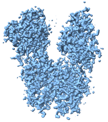
| タイトル | MicroED structure of an Aeropyrum pernix protoglobin mutant | |||||||||
 マップデータ マップデータ | ||||||||||
 試料 試料 |
| |||||||||
 キーワード キーワード | MicroED / protoglobin / directed evolution / METAL BINDING PROTEIN | |||||||||
| 機能・相同性 | Protoglobin / Globin-sensor domain / Protoglobin / Globin/Protoglobin / Globin-like superfamily / oxygen binding / heme binding / Protogloblin ApPgb 機能・相同性情報 機能・相同性情報 | |||||||||
| 生物種 |   Aeropyrum pernix (古細菌) Aeropyrum pernix (古細菌) | |||||||||
| 手法 | 電子線結晶学 / クライオ電子顕微鏡法 / 解像度: 2.1 Å | |||||||||
 データ登録者 データ登録者 | Danelius E / Gonen T / Unge JT | |||||||||
| 資金援助 |  米国, 2件 米国, 2件
| |||||||||
 引用 引用 | ジャーナル: J Am Chem Soc / 年: 2023 タイトル: MicroED Structure of a Protoglobin Reactive Carbene Intermediate. 著者: Emma Danelius / Nicholas J Porter / Johan Unge / Frances H Arnold / Tamir Gonen / 要旨: Microcrystal electron diffraction (MicroED) is an emerging technique that has shown great potential for describing new chemical and biological molecular structures. Several important structures of ...Microcrystal electron diffraction (MicroED) is an emerging technique that has shown great potential for describing new chemical and biological molecular structures. Several important structures of small molecules, natural products, and peptides have been determined using methods. However, only a couple of novel protein structures have thus far been derived by MicroED. Taking advantage of recent technological advances, including higher acceleration voltage and using a low-noise detector in counting mode, we have determined the first structure of an protoglobin (Pgb) variant by MicroED using an AlphaFold2 model for phasing. The structure revealed that mutations introduced during directed evolution enhance carbene transfer activity by reorienting an α helix of Pgb into a dynamic loop, making the catalytic active site more readily accessible. After exposing the tiny crystals to the substrate, we also trapped the reactive iron-carbenoid intermediate involved in this engineered Pgb's new-to-nature activity, a challenging carbene transfer from a diazirine via a putative metallo-carbene. The bound structure discloses how an enlarged active site pocket stabilizes the carbene bound to the heme iron and, presumably, the transition state for the formation of this key intermediate. This work demonstrates that improved MicroED technology and the advancement in protein structure prediction now enable investigation of structures that was previously beyond reach. | |||||||||
| 履歴 |
|
- 構造の表示
構造の表示
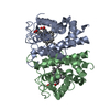
| 添付画像 |
|---|
- ダウンロードとリンク
-EMDBアーカイブ
| マップデータ | emd_28615.map.gz | 19.4 MB | EMDBマップデータ形式 | |
|---|---|---|---|---|
| ヘッダ (付随情報) | emd-28615-v30.xmlemd-28615.xml | 14.2 KB 14.2 KB | 表示 表示 | EMDBヘッダ |
| 画像 |  emd_28615.png emd_28615.png | 181.2 KB | ||
| Filedesc metadata | emd-28615.cif.gz | 5.9 KB | ||
| アーカイブディレクトリ |  http://ftp.pdbj.org/pub/emdb/structures/EMD-28615ftp://ftp.pdbj.org/pub/emdb/structures/EMD-28615 http://ftp.pdbj.org/pub/emdb/structures/EMD-28615ftp://ftp.pdbj.org/pub/emdb/structures/EMD-28615 | HTTPS FTP |
-検証レポート
| 文書・要旨 | emd_28615_validation.pdf.gz | 668.9 KB | 表示 | EMDB検証レポート |
|---|---|---|---|---|
| 文書・詳細版 | emd_28615_full_validation.pdf.gz | 668.5 KB | 表示 | |
| XML形式データ | emd_28615_validation.xml.gz | 4.5 KB | 表示 | |
| CIF形式データ | emd_28615_validation.cif.gz | 5 KB | 表示 | |
| アーカイブディレクトリ | https://ftp.pdbj.org/pub/emdb/validation_reports/EMD-28615ftp://ftp.pdbj.org/pub/emdb/validation_reports/EMD-28615 | HTTPS FTP |
-関連構造データ

-リンク
| EMDBのページ | EMDB (EBI/PDBe) / EMDataResource |
|---|---|
| 「今月の分子」の関連する項目 |


-マップ
| ファイル | ダウンロード / ファイル: emd_28615.map.gz / 形式: CCP4 / 大きさ: 25.4 MB / タイプ: IMAGE STORED AS FLOATING POINT NUMBER (4 BYTES) | ||||||||||||||||||||||||||||||||||||
|---|---|---|---|---|---|---|---|---|---|---|---|---|---|---|---|---|---|---|---|---|---|---|---|---|---|---|---|---|---|---|---|---|---|---|---|---|---|
| 投影像・断面図 | 画像のコントロール
画像は Spider により作成 これらの図は立方格子座標系で作成されたものです | ||||||||||||||||||||||||||||||||||||
| ボクセルのサイズ | X: 0.51372 Å / Y: 0.48558 Å / Z: 0.50459 Å | ||||||||||||||||||||||||||||||||||||
| 密度 |
| ||||||||||||||||||||||||||||||||||||
| 対称性 | 空間群: 1 | ||||||||||||||||||||||||||||||||||||
| 詳細 | EMDB XML:
|
 X (Sec.)
 Y (Row.)
 Z (Col.)


-添付データ
- 試料の構成要素
-全体 : Homotetramer of Aeropyrum pernix protoglobin (ApePgb) C45G, W59L,...
| 全体 | 名称: Homotetramer of Aeropyrum pernix protoglobin (ApePgb) C45G, W59L, Y60V, V63R, C102S, F145Q, I149L mutant |
|---|---|
| 要素 |
|
-超分子 #1: Homotetramer of Aeropyrum pernix protoglobin (ApePgb) C45G, W59L,...
| 超分子 | 名称: Homotetramer of Aeropyrum pernix protoglobin (ApePgb) C45G, W59L, Y60V, V63R, C102S, F145Q, I149L mutant タイプ: complex / ID: 1 / 親要素: 0 / 含まれる分子: #1 |
|---|---|
| 由来(天然) | 生物種: Aeropyrum pernix (古細菌) |
-分子 #1: Protogloblin ApPgb
| 分子 | 名称: Protogloblin ApPgb / タイプ: protein_or_peptide / ID: 1 / コピー数: 4 / 光学異性体: LEVO |
|---|---|
| 由来(天然) | 生物種: Aeropyrum pernix (古細菌) |
| 分子量 | 理論値: 22.187307 KDa |
| 組換発現 | 生物種:  |
| 配列 | 文字列: IPGYDYGRVE KSPITDLEFD LLKKTVMLGE KDVMYLKKAG DVLKDQVDEI LDLLVGWRAS NEHLIYYFSN PDTGEPIKEY LERVRARFG AWILDTTSRD YNREWLDYQY EVGLRHHRSK KGVTDGVRTV PHIPLRYLIA QIYPLTATIK PFLAKKGGSP E DIEGMYNA ...文字列: IPGYDYGRVE KSPITDLEFD LLKKTVMLGE KDVMYLKKAG DVLKDQVDEI LDLLVGWRAS NEHLIYYFSN PDTGEPIKEY LERVRARFG AWILDTTSRD YNREWLDYQY EVGLRHHRSK KGVTDGVRTV PHIPLRYLIA QIYPLTATIK PFLAKKGGSP E DIEGMYNA WFKSVVLQVA IWSHPYTKEN DW UniProtKB: Protogloblin ApPgb |
-分子 #2: PROTOPORPHYRIN IX CONTAINING FE
| 分子 | 名称: PROTOPORPHYRIN IX CONTAINING FE / タイプ: ligand / ID: 2 / コピー数: 4 / 式: HEM |
|---|---|
| 分子量 | 理論値: 616.487 Da |
| Chemical component information |  ChemComp-HEM: |
-分子 #3: IMIDAZOLE
| 分子 | 名称: IMIDAZOLE / タイプ: ligand / ID: 3 / コピー数: 15 / 式: IMD |
|---|---|
| 分子量 | 理論値: 69.085 Da |
| Chemical component information |  ChemComp-IMD: |
-分子 #4: FE (III) ION
| 分子 | 名称: FE (III) ION / タイプ: ligand / ID: 4 / コピー数: 4 / 式: FE |
|---|---|
| 分子量 | 理論値: 55.845 Da |
-分子 #5: water
| 分子 | 名称: water / タイプ: ligand / ID: 5 / コピー数: 308 / 式: HOH |
|---|---|
| 分子量 | 理論値: 18.015 Da |
| Chemical component information |  ChemComp-HOH: |
-実験情報
-構造解析
| 手法 | クライオ電子顕微鏡法 |
|---|---|
 解析 解析 | 電子線結晶学 |
| 試料の集合状態 | 3D array |
-試料調製
| 濃度 | 20 mg/mL |
|---|---|
| 緩衝液 | pH: 8 |
| グリッド | モデル: Quantifoil R2/2 / 材質: COPPER / メッシュ: 200 / 支持フィルム - 材質: CARBON / 支持フィルム - トポロジー: HOLEY / 前処理 - タイプ: GLOW DISCHARGE / 前処理 - 時間: 30 sec. / 前処理 - 雰囲気: OTHER |
| 凍結 | 凍結剤: ETHANE / チャンバー内湿度: 90 % / チャンバー内温度: 277 K / 装置: LEICA EM GP |
- 電子顕微鏡法
| 顕微鏡 | FEI TITAN KRIOS |
|---|---|
| 温度 | 最低: 77.0 K / 最高: 90.0 K |
| 撮影 | フィルム・検出器のモデル: FEI FALCON IV (4k x 4k) 検出モード: COUNTING / デジタル化 - サイズ - 横: 4096 pixel / デジタル化 - サイズ - 縦: 4096 pixel / 平均電子線量: 0.00025 e/Å2 |
| 電子線 | 加速電圧: 300 kV / 電子線源:  FIELD EMISSION GUN FIELD EMISSION GUN |
| 電子光学系 | C2レンズ絞り径: 50.0 µm / 照射モード: OTHER / 撮影モード: DIFFRACTION / Cs: 2.7 mm / 最大 デフォーカス(公称値): 0.0 µm / 最小 デフォーカス(公称値): 0.0 µm / カメラ長: 2460 mm |
| 試料ステージ | 試料ホルダーモデル: FEI TITAN KRIOS AUTOGRID HOLDER ホルダー冷却材: NITROGEN |
| 実験機器 |  モデル: Titan Krios / 画像提供: FEI Company |
-画像解析
| 最終 再構成 | 解像度のタイプ: BY AUTHOR / 解像度: 2.1 Å / 解像度の算出法: DIFFRACTION PATTERN/LAYERLINES / ソフトウェア - 名称: PHENIX (ver. 1.20.1-4487-000) |
|---|---|
| Crystallography statistics | Number intensities measured: 33826 / Number structure factors: 33826 / Fourier space coverage: 72 / R merge: 0.43 / Overall phase residual: 0 / High resolution: 2.1 Å / 殻 - Shell ID: 1 / 殻 - High resolution: 2.1 Å / 殻 - Low resolution: 24.27 Å / 殻 - Number structure factors: 33826 / 殻 - Phase residual: 21.11 / 殻 - Fourier space coverage: 73.1 / 殻 - Multiplicity: 4.1 |
-原子モデル構築 1
| 精密化 | 空間: RECIPROCAL / プロトコル: OTHER / 温度因子: 26.51 / 当てはまり具合の基準: maximum likelihood |
|---|---|
| 得られたモデル | 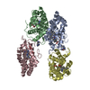 PDB-8eum: |
