National Institutes of Health/National Institute of General Medical Sciences (NIH/NIGMS)
1DP2GM146250
米国
The Vallee Foundation Inc.
David and Lucile Packard Foundation
米国
National Institutes of Health/National Institute of General Medical Sciences (NIH/NIGMS)
1DP2GM137415
米国
National Institutes of Health/National Institute of General Medical Sciences (NIH/NIGMS)
F32GM133063
米国
American Heart Association
287375208
米国
引用
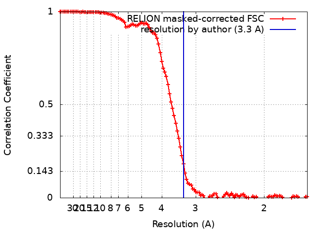
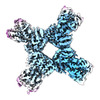
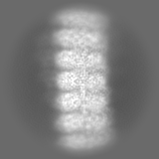
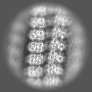
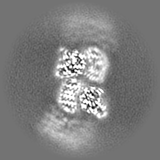
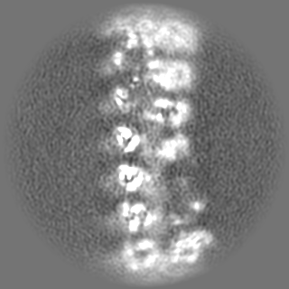
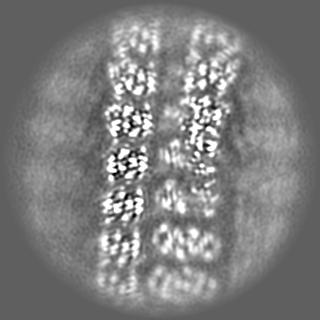
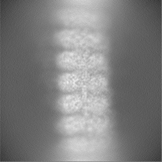
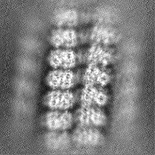
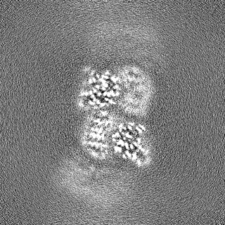
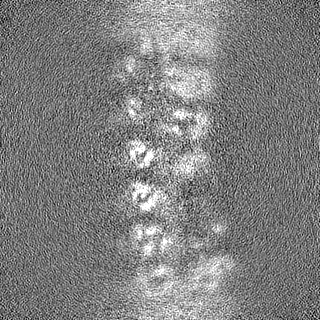
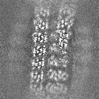
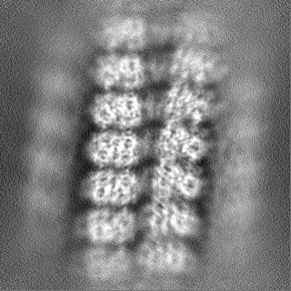
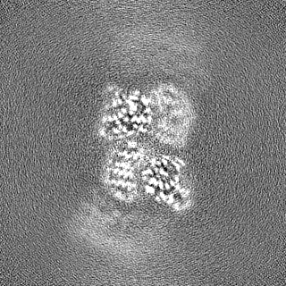
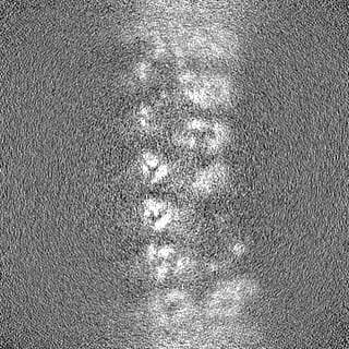
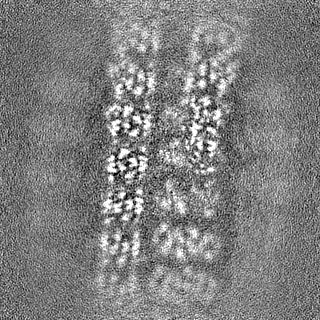
ジャーナル: Nature / 年: 2022 タイトル: Cryo-EM structure of an active bacterial TIR-STING filament complex. 著者: Benjamin R Morehouse / Matthew C J Yip / Alexander F A Keszei / Nora K McNamara-Bordewick / Sichen Shao / Philip J Kranzusch / 要旨: Stimulator of interferon genes (STING) is an antiviral signalling protein that is broadly conserved in both innate immunity in animals and phage defence in prokaryotes. Activation of STING requires ...Stimulator of interferon genes (STING) is an antiviral signalling protein that is broadly conserved in both innate immunity in animals and phage defence in prokaryotes. Activation of STING requires its assembly into an oligomeric filament structure through binding of a cyclic dinucleotide, but the molecular basis of STING filament assembly and extension remains unknown. Here we use cryogenic electron microscopy to determine the structure of the active Toll/interleukin-1 receptor (TIR)-STING filament complex from a Sphingobacterium faecium cyclic-oligonucleotide-based antiphage signalling system (CBASS) defence operon. Bacterial TIR-STING filament formation is driven by STING interfaces that become exposed on high-affinity recognition of the cognate cyclic dinucleotide signal c-di-GMP. Repeating dimeric STING units stack laterally head-to-head through surface interfaces, which are also essential for human STING tetramer formation and downstream immune signalling in mammals. The active bacterial TIR-STING structure reveals further cross-filament contacts that brace the assembly and coordinate packing of the associated TIR NADase effector domains at the base of the filament to drive NAD hydrolysis. STING interface and cross-filament contacts are essential for cell growth arrest in vivo and reveal a stepwise mechanism of activation whereby STING filament assembly is required for subsequent effector activation. Our results define the structural basis of STING filament formation in prokaryotic antiviral signalling.
 ムービー
ムービー コントローラー


 データを開く
 基本情報
基本情報
 マップデータ
 試料
試料 キーワード
キーワード 機能・相同性情報
 Sphingobacterium faecium (バクテリア)
 データ登録者
 米国, 11件
  引用
引用 構造の表示
構造の表示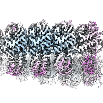
 ダウンロードとリンク
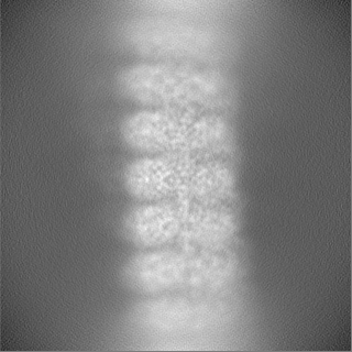
 emd_26617.png
 http://ftp.pdbj.org/pub/emdb/structures/EMD-26617


 Z
Z Y
Y X
X


 試料の構成要素
 解析
解析 電子顕微鏡法
 FIELD EMISSION GUN