Movie
Movie Controller

[English] 日本語
 Yorodumi
- EMDB-24765: Cryo-EM map of the phage AR9 non-virion RNA polymerase holoenzyme -
+ Open data

- Basic information
| Entry |  | |||||||||
|---|---|---|---|---|---|---|---|---|---|---|
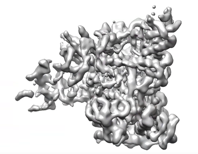
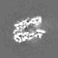
| Title | Cryo-EM map of the phage AR9 non-virion RNA polymerase holoenzyme | |||||||||
 Map data Map data | Cryo-EM map of the phage AR9 non-virion RNA polymerase holoenzyme | |||||||||
 Sample Sample |
| |||||||||
| Function / homology |  Function and homology information Function and homology informationRNA polymerase II activity / DNA-directed RNA polymerase complex / ribonucleoside binding / DNA-directed 5'-3' RNA polymerase activity / DNA-directed RNA polymerase / DNA-templated transcription / DNA binding / metal ion binding Similarity search - Function | |||||||||
| Biological species |  Bacillus phage AR9 (virus) Bacillus phage AR9 (virus) | |||||||||
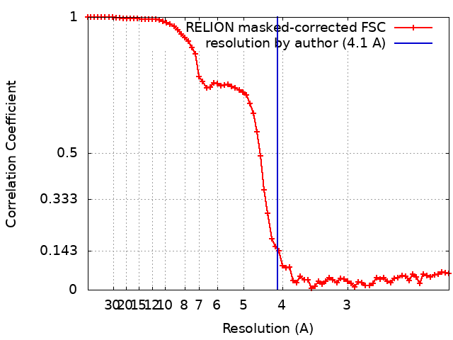
| Method | single particle reconstruction / cryo EM / Resolution: 4.1 Å | |||||||||
 Authors Authors | Fraser A / Leiman PG / Sokolova ML | |||||||||
| Funding support | 1 items
| |||||||||
 Citation Citation | Journal: Nat Commun / Year: 2022 Title: Structural basis of template strand deoxyuridine promoter recognition by a viral RNA polymerase. Authors: Alec Fraser / Maria L Sokolova / Arina V Drobysheva / Julia V Gordeeva / Sergei Borukhov / John Jumper / Konstantin V Severinov / Petr G Leiman /    Abstract: Recognition of promoters in bacterial RNA polymerases (RNAPs) is controlled by sigma subunits. The key sequence motif recognized by the sigma, the -10 promoter element, is located in the non-template ...Recognition of promoters in bacterial RNA polymerases (RNAPs) is controlled by sigma subunits. The key sequence motif recognized by the sigma, the -10 promoter element, is located in the non-template strand of the double-stranded DNA molecule ~10 nucleotides upstream of the transcription start site. Here, we explain the mechanism by which the phage AR9 non-virion RNAP (nvRNAP), a bacterial RNAP homolog, recognizes the -10 element of its deoxyuridine-containing promoter in the template strand. The AR9 sigma-like subunit, the nvRNAP enzyme core, and the template strand together form two nucleotide base-accepting pockets whose shapes dictate the requirement for the conserved deoxyuridines. A single amino acid substitution in the AR9 sigma-like subunit allows one of these pockets to accept a thymine thus expanding the promoter consensus. Our work demonstrates the extent to which viruses can evolve host-derived multisubunit enzymes to make transcription of their own genes independent of the host. | |||||||||
| History |
|
- Structure visualization
| Supplemental images |
|---|
- Downloads & links
-EMDB archive
| Map data | emd_24765.map.gz | 28.1 MB | EMDB map data format | |
|---|---|---|---|---|
| Header (meta data) | emd-24765-v30.xmlemd-24765.xml | 12.3 KB 12.3 KB | Display Display | EMDB header |
| FSC (resolution estimation) | emd_24765_fsc.xml | 7.2 KB | Display | FSC data file |
| Images |  emd_24765.png emd_24765.png | 114.8 KB | ||
| Others | emd_24765_half_map_1.map.gzemd_24765_half_map_2.map.gz | 22.9 MB 22.9 MB | ||
| Archive directory |  http://ftp.pdbj.org/pub/emdb/structures/EMD-24765ftp://ftp.pdbj.org/pub/emdb/structures/EMD-24765 http://ftp.pdbj.org/pub/emdb/structures/EMD-24765ftp://ftp.pdbj.org/pub/emdb/structures/EMD-24765 | HTTPS FTP |
-Validation report
| Summary document | emd_24765_validation.pdf.gz | 651.5 KB | Display | EMDB validaton report |
|---|---|---|---|---|
| Full document | emd_24765_full_validation.pdf.gz | 651.1 KB | Display | |
| Data in XML | emd_24765_validation.xml.gz | 13.1 KB | Display | |
| Data in CIF | emd_24765_validation.cif.gz | 17.9 KB | Display | |
| Arichive directory | https://ftp.pdbj.org/pub/emdb/validation_reports/EMD-24765ftp://ftp.pdbj.org/pub/emdb/validation_reports/EMD-24765 | HTTPS FTP |
-Related structure data
| Related structure data | 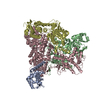 7um1MC 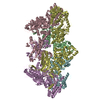 7s00C 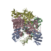 7s01C 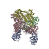 7um0C M: atomic model generated by this map C: citing same article ( |
|---|---|
| Similar structure data |

-Links
| EMDB pages | EMDB (EBI/PDBe) / EMDataResource |
|---|---|
| Related items in Molecule of the Month |

-Map
| File | Download / File: emd_24765.map.gz / Format: CCP4 / Size: 30.5 MB / Type: IMAGE STORED AS FLOATING POINT NUMBER (4 BYTES) | ||||||||||||||||||||||||||||||||||||
|---|---|---|---|---|---|---|---|---|---|---|---|---|---|---|---|---|---|---|---|---|---|---|---|---|---|---|---|---|---|---|---|---|---|---|---|---|---|
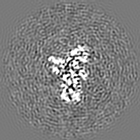
| Annotation | Cryo-EM map of the phage AR9 non-virion RNA polymerase holoenzyme | ||||||||||||||||||||||||||||||||||||
| Projections & slices | Image control
Images are generated by Spider. | ||||||||||||||||||||||||||||||||||||
| Voxel size | X=Y=Z: 1.08 Å | ||||||||||||||||||||||||||||||||||||
| Density |
| ||||||||||||||||||||||||||||||||||||
| Symmetry | Space group: 1 | ||||||||||||||||||||||||||||||||||||
| Details | EMDB XML:
|
 Z (Sec.)
 Y (Row.)
 X (Col.)


-Supplemental data
-Half map: Half map A
| File | emd_24765_half_map_1.map | ||||||||||||
|---|---|---|---|---|---|---|---|---|---|---|---|---|---|
| Annotation | Half map A | ||||||||||||
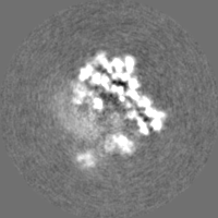
| Projections & Slices |
| ||||||||||||
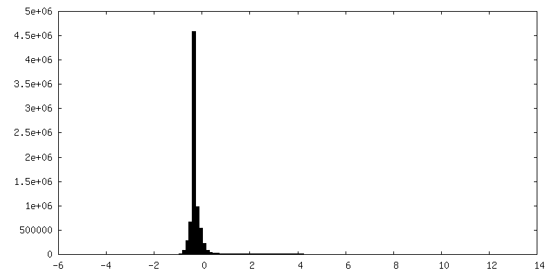
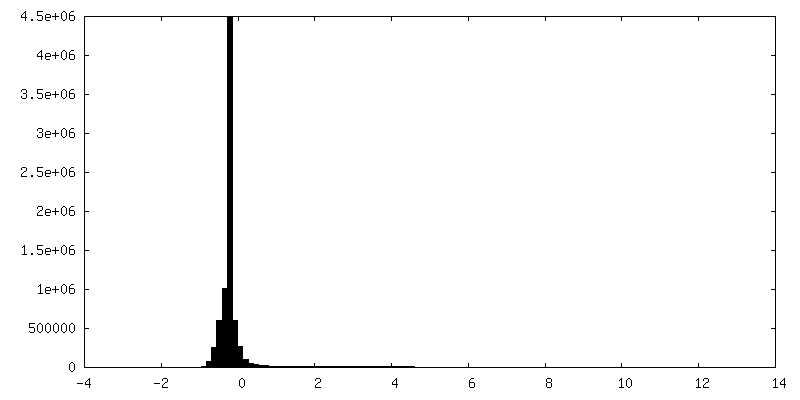
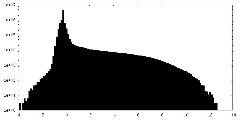
| Density Histograms |

-Half map: Half map B
| File | emd_24765_half_map_2.map | ||||||||||||
|---|---|---|---|---|---|---|---|---|---|---|---|---|---|
| Annotation | Half map B | ||||||||||||
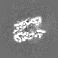
| Projections & Slices |
| ||||||||||||
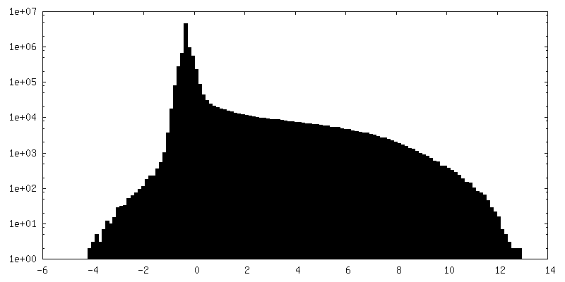
| Density Histograms |

- Sample components
-Entire : Bacteriophage AR9 non-virion RNA polymerase holoenzyme
| Entire | Name: Bacteriophage AR9 non-virion RNA polymerase holoenzyme |
|---|---|
| Components |
|
-Supramolecule #1: Bacteriophage AR9 non-virion RNA polymerase holoenzyme
| Supramolecule | Name: Bacteriophage AR9 non-virion RNA polymerase holoenzyme type: complex / ID: 1 / Chimera: Yes / Parent: 0 / Macromolecule list: #1 |
|---|---|
| Source (natural) | Organism: Bacillus phage AR9 (virus) |
-Experimental details
-Structure determination
| Method | cryo EM |
|---|---|
 Processing Processing | single particle reconstruction |
| Aggregation state | particle |
-Sample preparation
| Concentration | 20 mg/mL |
|---|---|
| Buffer | pH: 6.8 |
| Vitrification | Cryogen name: ETHANE |
- Electron microscopy
| Microscope | FEI TITAN KRIOS |
|---|---|
| Image recording | Film or detector model: GATAN K2 SUMMIT (4k x 4k) / Average electron dose: 43.2 e/Å2 |
| Electron beam | Acceleration voltage: 300 kV / Electron source:  FIELD EMISSION GUN FIELD EMISSION GUN |
| Electron optics | Illumination mode: FLOOD BEAM / Imaging mode: BRIGHT FIELD / Nominal defocus max: 4.0 µm / Nominal defocus min: 1.0 µm |
| Experimental equipment |  Model: Titan Krios / Image courtesy: FEI Company |