 ムービー
ムービー コントローラー


+ データを開く

- 基本情報
基本情報
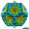
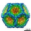
| 登録情報 | データベース: EMDB / ID: EMD-2364 | |||||||||
|---|---|---|---|---|---|---|---|---|---|---|
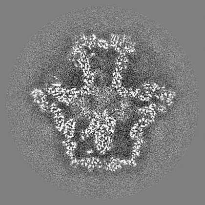
| タイトル | CryoEM reconstruction of the bacteriophage phi6 procapsid to the near-atomic resolution | |||||||||
 マップデータ マップデータ | Reconstruction of the wildtype P1247 procapsid of bacteriophage phi6 | |||||||||
 試料 試料 |
| |||||||||
 キーワード キーワード | Bacteriophage phi6 / Cystoviridae / capsid structure / capsid expansion / segmented genome / conformational change / RNA packaging | |||||||||
| 機能・相同性 | : / Major inner capsid protein P1 / T=2 icosahedral viral capsid / viral inner capsid / viral nucleocapsid / RNA binding / identical protein binding / Major inner protein P1 機能・相同性情報 機能・相同性情報 | |||||||||
| 生物種 |  Pseudomonas phage phi6 (ファージ) Pseudomonas phage phi6 (ファージ) | |||||||||
| 手法 | 単粒子再構成法 / クライオ電子顕微鏡法 / 解像度: 4.4 Å | |||||||||
 データ登録者 データ登録者 | Nemecek D / Boura E / Wu W / Cheng N / Plevka P / Qiao J / Mindich L / Heymann JB / Hurley JH / Steven AC | |||||||||
 引用 引用 | ジャーナル: Structure / 年: 2013 タイトル: Subunit folds and maturation pathway of a dsRNA virus capsid. 著者: Daniel Nemecek / Evzen Boura / Weimin Wu / Naiqian Cheng / Pavel Plevka / Jian Qiao / Leonard Mindich / J Bernard Heymann / James H Hurley / Alasdair C Steven /  要旨: The cystovirus ϕ6 shares several distinct features with other double-stranded RNA (dsRNA) viruses, including the human pathogen, rotavirus: segmented genomes, nonequivalent packing of 120 subunits ...The cystovirus ϕ6 shares several distinct features with other double-stranded RNA (dsRNA) viruses, including the human pathogen, rotavirus: segmented genomes, nonequivalent packing of 120 subunits in its icosahedral capsid, and capsids as compartments for transcription and replication. ϕ6 assembles as a dodecahedral procapsid that undergoes major conformational changes as it matures into the spherical capsid. We determined the crystal structure of the capsid protein, P1, revealing a flattened trapezoid subunit with an α-helical fold. We also solved the procapsid with cryo-electron microscopy to comparable resolution. Fitting the crystal structure into the procapsid disclosed substantial conformational differences between the two P1 conformers. Maturation via two intermediate states involves remodeling on a similar scale, besides huge rigid-body rotations. The capsid structure and its stepwise maturation that is coupled to sequential packaging of three RNA segments sets the cystoviruses apart from other dsRNA viruses as a dynamic molecular machine. | |||||||||
| 履歴 |
|
- 構造の表示
構造の表示
| ムービー |
ムービービューア |
|---|---|
| 構造ビューア | EMマップ: SurfViewMolmilJmol/JSmol |
| 添付画像 |

- ダウンロードとリンク
-EMDBアーカイブ
| マップデータ | emd_2364.map.gz | 265.9 MB | EMDBマップデータ形式 | |
|---|---|---|---|---|
| ヘッダ (付随情報) | emd-2364-v30.xmlemd-2364.xml | 15.1 KB 15.1 KB | 表示 表示 | EMDBヘッダ |
| FSC (解像度算出) | emd_2364_fsc.xml | 15.5 KB | 表示 | FSCデータファイル |
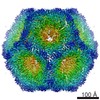
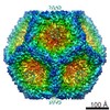
| 画像 | 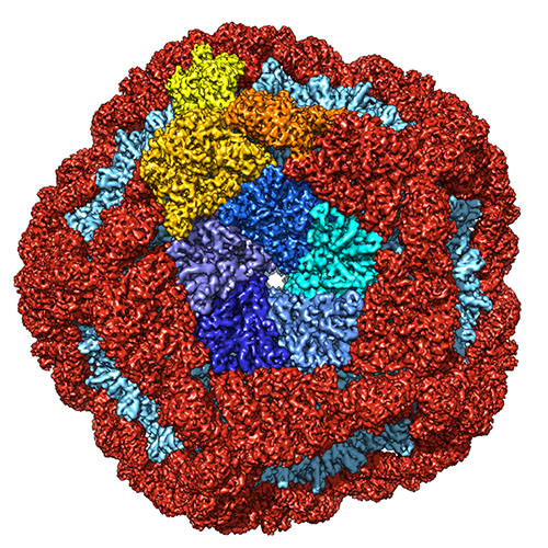 emd_2364.jpg emd_2364.jpg | 193.8 KB | ||
| マスクデータ | emd_2364_msk_1.mapemd_2364_msk_2.map | 70.7 MB 70.7 MB | マスクマップ | |
| アーカイブディレクトリ |  http://ftp.pdbj.org/pub/emdb/structures/EMD-2364ftp://ftp.pdbj.org/pub/emdb/structures/EMD-2364 http://ftp.pdbj.org/pub/emdb/structures/EMD-2364ftp://ftp.pdbj.org/pub/emdb/structures/EMD-2364 | HTTPS FTP |
-関連構造データ

-リンク
| EMDBのページ | EMDB (EBI/PDBe) / EMDataResource |
|---|---|
| 「今月の分子」の関連する項目 |

-マップ
| ファイル | ダウンロード / ファイル: emd_2364.map.gz / 形式: CCP4 / 大きさ: 276 MB / タイプ: IMAGE STORED AS FLOATING POINT NUMBER (4 BYTES) | ||||||||||||||||||||||||||||||||||||||||||||||||||||||||||||||||||||
|---|---|---|---|---|---|---|---|---|---|---|---|---|---|---|---|---|---|---|---|---|---|---|---|---|---|---|---|---|---|---|---|---|---|---|---|---|---|---|---|---|---|---|---|---|---|---|---|---|---|---|---|---|---|---|---|---|---|---|---|---|---|---|---|---|---|---|---|---|---|
| 注釈 | Reconstruction of the wildtype P1247 procapsid of bacteriophage phi6 | ||||||||||||||||||||||||||||||||||||||||||||||||||||||||||||||||||||
| 投影像・断面図 | 画像のコントロール
画像は Spider により作成 | ||||||||||||||||||||||||||||||||||||||||||||||||||||||||||||||||||||
| ボクセルのサイズ | X=Y=Z: 1.397 Å | ||||||||||||||||||||||||||||||||||||||||||||||||||||||||||||||||||||
| 密度 |
| ||||||||||||||||||||||||||||||||||||||||||||||||||||||||||||||||||||
| 対称性 | 空間群: 1 | ||||||||||||||||||||||||||||||||||||||||||||||||||||||||||||||||||||
| 詳細 | EMDB XML:
CCP4マップ ヘッダ情報:
| ||||||||||||||||||||||||||||||||||||||||||||||||||||||||||||||||||||
 Z (Sec.)
 Y (Row.)
 X (Col.)


-添付データ
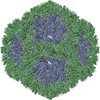
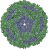
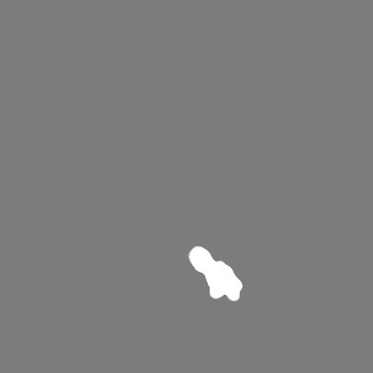
-セグメンテーションマップ: This mask represents the P1B subunit
| 注釈 | This mask represents the P1B subunit | ||||||||||||
|---|---|---|---|---|---|---|---|---|---|---|---|---|---|
| ファイル | emd_2364_msk_1.map | ||||||||||||
| 投影像・断面図 |
| ||||||||||||
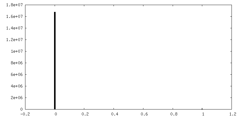
| 密度ヒストグラム |

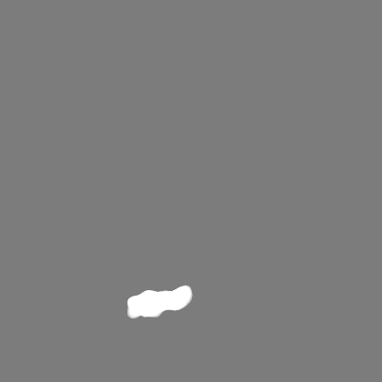


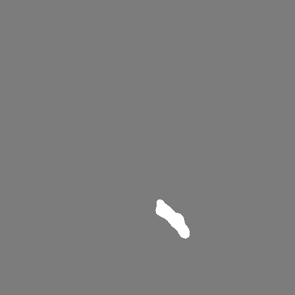
-セグメンテーションマップ: This mask represents the P1A subunit
| 注釈 | This mask represents the P1A subunit | ||||||||||||
|---|---|---|---|---|---|---|---|---|---|---|---|---|---|
| ファイル | emd_2364_msk_2.map | ||||||||||||
| 投影像・断面図 |
| ||||||||||||
| 密度ヒストグラム |


- 試料の構成要素
-全体 : Wildtype P1247 procapsid of bacteriophage phi6
| 全体 | 名称: Wildtype P1247 procapsid of bacteriophage phi6 |
|---|---|
| 要素 |
|
-超分子 #1000: Wildtype P1247 procapsid of bacteriophage phi6
| 超分子 | 名称: Wildtype P1247 procapsid of bacteriophage phi6 / タイプ: sample / ID: 1000 / 集合状態: icosahedral shell with accessory proteins / Number unique components: 4 |
|---|---|
| 分子量 | 理論値: 12.6 MDa |
-超分子 #1: Pseudomonas phage phi6
| 超分子 | 名称: Pseudomonas phage phi6 / タイプ: virus / ID: 1 / Name.synonym: bacteriophage phi-6 / NCBI-ID: 10879 / 生物種: Pseudomonas phage phi6 / ウイルスタイプ: VIRION / ウイルス・単離状態: SPECIES / ウイルス・エンベロープ: No / ウイルス・中空状態: Yes / Syn species name: bacteriophage phi-6 |
|---|---|
| 宿主 | 生物種:  Pseudomonas syringae (バクテリア) / 別称: BACTERIA(EUBACTERIA) Pseudomonas syringae (バクテリア) / 別称: BACTERIA(EUBACTERIA) |
| Host system | 生物種: |
| 分子量 | 理論値: 12.6 MDa |
| ウイルス殻 | Shell ID: 1 / 名称: P1247 / 直径: 450 Å / T番号(三角分割数): 2 |
-実験情報
-構造解析
| 手法 | クライオ電子顕微鏡法 |
|---|---|
 解析 解析 | 単粒子再構成法 |
| 試料の集合状態 | particle |
-試料調製
| 濃度 | 10 mg/mL |
|---|---|
| 緩衝液 | pH: 8 / 詳細: 10 mM potassium phosphate, 5 mM MgCl2 |
| グリッド | 詳細: 400 mesh C-flat holey carbon grid |
| 凍結 | 凍結剤: ETHANE / チャンバー内湿度: 80 % / チャンバー内温度: 100 K / 装置: FEI VITROBOT MARK I / 手法: Blot for 2 seconds before plunging. |
- 電子顕微鏡法
| 顕微鏡 | FEI TITAN KRIOS |
|---|---|
| 日付 | 2011年11月20日 |
| 撮影 | カテゴリ: FILM / フィルム・検出器のモデル: KODAK SO-163 FILM デジタル化 - スキャナー: NIKON SUPER COOLSCAN 9000 デジタル化 - サンプリング間隔: 6.35 µm / 実像数: 220 / 平均電子線量: 15 e/Å2 / ビット/ピクセル: 16 |
| Tilt angle min | 0 |
| Tilt angle max | 0 |
| 電子線 | 加速電圧: 300 kV / 電子線源:  FIELD EMISSION GUN FIELD EMISSION GUN |
| 電子光学系 | 倍率(補正後): 44739 / 照射モード: FLOOD BEAM / 撮影モード: BRIGHT FIELD / Cs: 2.7 mm / 最大 デフォーカス(公称値): 2.2 µm / 最小 デフォーカス(公称値): 0.8 µm / 倍率(公称値): 46000 |
| 試料ステージ | 試料ホルダーモデル: FEI TITAN KRIOS AUTOGRID HOLDER |
| 実験機器 |  モデル: Titan Krios / 画像提供: FEI Company |
-画像解析
| 詳細 | The particles were selected using e2boxer (EMAN) and manually pruned in bshow (BSOFT). The initial model was taken from our previous reconstruction at 7A resolution (EMD-2341). The final structure was reconstructed in EMAN. |
|---|---|
| CTF補正 | 詳細: Particles from each micrograph |
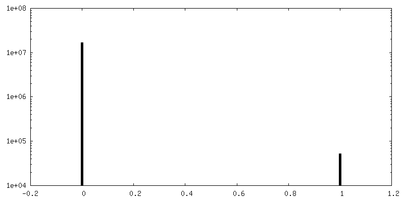
| 最終 再構成 | 想定した対称性 - 点群: I (正20面体型対称) / 解像度のタイプ: BY AUTHOR / 解像度: 4.4 Å / 解像度の算出法: FSC 0.5 CUT-OFF / ソフトウェア - 名称: EMAN, BSOFT / 使用した粒子像数: 18326 |
| FSC曲線 (解像度の算出) | 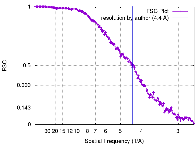 |
-原子モデル構築 1
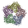
| 初期モデル | PDB ID: Chain - Chain ID: A |
|---|---|
| ソフトウェア | 名称: Chimera, MDFF, Coot |
| 詳細 | the P1 crystal structure was rigid body-fitted into the procapsid map using Chimera. Regions where the crystal structure deviated significantly from the EM density were roughly adjusted in Coot and then the P1A and P1B structures were flexibly fitted using the MDFF package. Finally, both structures were visually inspected and refined in Coot. |
| 精密化 | 空間: REAL / プロトコル: FLEXIBLE FIT |
| 得られたモデル |  PDB-4btg: |
