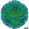
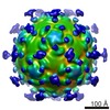
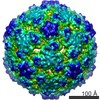
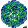
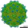
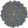
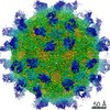
Journal: PLoS One / Year: 2013 Title: 3.5Å cryoEM structure of hepatitis B virus core assembled from full-length core protein. Authors: Xuekui Yu / Lei Jin / Jonathan Jih / Chiaho Shih / Z Hong Zhou / Abstract: The capsid shell of infectious hepatitis B virus (HBV) is composed of 240 copies of a single protein called HBV core antigen (HBc). An atomic model of a core assembled from truncated HBc was ...The capsid shell of infectious hepatitis B virus (HBV) is composed of 240 copies of a single protein called HBV core antigen (HBc). An atomic model of a core assembled from truncated HBc was determined previously by X-ray crystallography. In an attempt to obtain atomic structural information of HBV core in a near native, non-crystalline environment, we reconstructed a 3.5Å-resolution structure of a recombinant core assembled from full-length HBc by cryo electron microscopy (cryoEM) and derived an atomic model. The structure shows that the 240 molecules of full-length HBc form a core with two layers. The outer layer, composed of the N-terminal assembly domain, is similar to the crystal structure of the truncated HBc, but has three differences. First, unlike the crystal structure, our cryoEM structure shows no disulfide bond between the Cys61 residues of the two subunits within the dimer building block, indicating such bond is not required for core formation. Second, our cryoEM structure reveals up to four more residues in the linker region (amino acids 140-149). Third, the loops in the cryoEM structures containing this linker region in subunits B and C are oriented differently (~30° and ~90°) from their counterparts in the crystal structure. The inner layer, composed of the C-terminal arginine-rich domain (ARD) and the ARD-bound RNAs, is partially-ordered and connected with the outer layer through linkers positioned around the two-fold axes. Weak densities emanate from the rims of positively charged channels through the icosahedral three-fold and local three-fold axes. We attribute these densities to the exposed portions of some ARDs, thus explaining ARD's accessibility by proteases and antibodies. Our data supports a role of ARD in mediating communication between inside and outside of the core during HBV maturation and envelopment.
History
Deposition
Jan 11, 2013
-
Header (metadata) release
Jan 30, 2013
-
Map release
Oct 2, 2013
-
Update
Oct 2, 2013
-
Current status
Oct 2, 2013
Processing site: PDBe / Status: Released
-
Structure visualization
Movie
Surface view with section colored by density value
In the structure databanks used in Yorodumi, some data are registered as the other names, "COVID-19 virus" and "2019-nCoV". Here are the details of the virus and the list of structure data.
Jan 31, 2019. EMDB accession codes are about to change! (news from PDBe EMDB page)
EMDB accession codes are about to change! (news from PDBe EMDB page)
The allocation of 4 digits for EMDB accession codes will soon come to an end. Whilst these codes will remain in use, new EMDB accession codes will include an additional digit and will expand incrementally as the available range of codes is exhausted. The current 4-digit format prefixed with “EMD-” (i.e. EMD-XXXX) will advance to a 5-digit format (i.e. EMD-XXXXX), and so on. It is currently estimated that the 4-digit codes will be depleted around Spring 2019, at which point the 5-digit format will come into force.
The EM Navigator/Yorodumi systems omit the EMD- prefix.
Related info.:Q: What is EMD? / ID/Accession-code notation in Yorodumi/EM Navigator
Yorodumi is a browser for structure data from EMDB, PDB, SASBDB, etc.
This page is also the successor to EM Navigator detail page, and also detail information page/front-end page for Omokage search.
The word "yorodu" (or yorozu) is an old Japanese word meaning "ten thousand". "mi" (miru) is to see.
Related info.:EMDB / PDB / SASBDB / Comparison of 3 databanks / Yorodumi Search / Aug 31, 2016. New EM Navigator & Yorodumi / Yorodumi Papers / Jmol/JSmol / Function and homology information / Changes in new EM Navigator and Yorodumi
 Movie
Movie Controller

 Yorodumi
 Open data
 Basic information
 Map data
 Sample
 Keywords
 Function and homology information
 Hepatitis B virus
 Authors
 Citation
 Structure visualization
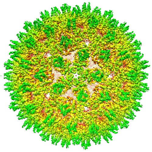
 Downloads & links
 EMD-2278-HBV.jpg
 http://ftp.pdbj.org/pub/emdb/structures/EMD-2278


 Z (Sec.)
 Y (Row.)
 X (Col.)


 Sample components
 Homo sapiens (human) / synonym: VERTEBRATES
 Processing
 Electron microscopy
 FIELD EMISSION GUN
