Movie
Movie Controller

[English] 日本語
 Yorodumi
- EMDB-22198: Cryo-EM Structure of K63 Ubiquitinated Yeast Translocating Riboso... -
+ Open data

- Basic information
| Entry | Database: EMDB / ID: EMD-22198 | |||||||||
|---|---|---|---|---|---|---|---|---|---|---|
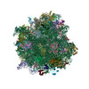
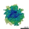
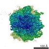
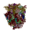
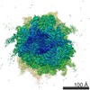
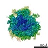
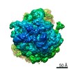
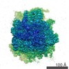
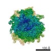
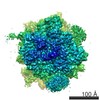
| Title | Cryo-EM Structure of K63 Ubiquitinated Yeast Translocating Ribosome under Oxidative Stress | |||||||||
 Map data Map data | ||||||||||
 Sample Sample |
| |||||||||
 Keywords Keywords | K63 ubiquitin / ribosome / oxidative stress / translation | |||||||||
| Function / homology |  Function and homology information Function and homology informationregulation of amino acid metabolic process / negative regulation of glucose mediated signaling pathway / mTORC1-mediated signalling / Protein hydroxylation / ribosome-associated ubiquitin-dependent protein catabolic process / GDP-dissociation inhibitor activity / regulation of polysaccharide biosynthetic process / Formation of the ternary complex, and subsequently, the 43S complex / Translation initiation complex formation / Ribosomal scanning and start codon recognition ...regulation of amino acid metabolic process / negative regulation of glucose mediated signaling pathway / mTORC1-mediated signalling / Protein hydroxylation / ribosome-associated ubiquitin-dependent protein catabolic process / GDP-dissociation inhibitor activity / regulation of polysaccharide biosynthetic process / Formation of the ternary complex, and subsequently, the 43S complex / Translation initiation complex formation / Ribosomal scanning and start codon recognition / nonfunctional rRNA decay / transporter complex / cleavage in ITS2 between 5.8S rRNA and LSU-rRNA of tricistronic rRNA transcript (SSU-rRNA, 5.8S rRNA, LSU-rRNA) / response to cycloheximide / Major pathway of rRNA processing in the nucleolus and cytosol / mRNA destabilization / lipopolysaccharide transport / SRP-dependent cotranslational protein targeting to membrane / GTP hydrolysis and joining of the 60S ribosomal subunit / Nonsense Mediated Decay (NMD) independent of the Exon Junction Complex (EJC) / Nonsense Mediated Decay (NMD) enhanced by the Exon Junction Complex (EJC) / negative regulation of translational frameshifting / Formation of a pool of free 40S subunits / positive regulation of protein kinase activity / preribosome, large subunit precursor / L13a-mediated translational silencing of Ceruloplasmin expression / Gram-negative-bacterium-type cell outer membrane assembly / endonucleolytic cleavage to generate mature 3'-end of SSU-rRNA from (SSU-rRNA, 5.8S rRNA, LSU-rRNA) / G-protein alpha-subunit binding / 90S preribosome / ribosomal subunit export from nucleus / regulation of translational fidelity / endonucleolytic cleavage in ITS1 to separate SSU-rRNA from 5.8S rRNA and LSU-rRNA from tricistronic rRNA transcript (SSU-rRNA, 5.8S rRNA, LSU-rRNA) / protein-RNA complex assembly / maturation of LSU-rRNA / translation regulator activity / rescue of stalled ribosome / protein kinase C binding / cellular response to amino acid starvation / maturation of LSU-rRNA from tricistronic rRNA transcript (SSU-rRNA, 5.8S rRNA, LSU-rRNA) / ribosomal large subunit biogenesis / positive regulation of apoptotic signaling pathway / maturation of SSU-rRNA from tricistronic rRNA transcript (SSU-rRNA, 5.8S rRNA, LSU-rRNA) / maturation of SSU-rRNA / macroautophagy / small-subunit processome / translational initiation / maintenance of translational fidelity / cell outer membrane / cytoplasmic stress granule / rRNA processing / ribosome biogenesis / ribosome binding / ribosomal small subunit biogenesis / ribosomal small subunit assembly / small ribosomal subunit / 5S rRNA binding / small ribosomal subunit rRNA binding / ribosomal large subunit assembly / cytosolic small ribosomal subunit / large ribosomal subunit rRNA binding / cytosolic large ribosomal subunit / cytoplasmic translation / negative regulation of translation / rRNA binding / structural constituent of ribosome / ribosome / translation / G protein-coupled receptor signaling pathway / negative regulation of gene expression / response to antibiotic / mRNA binding / nucleolus / mitochondrion / RNA binding / zinc ion binding / nucleoplasm / metal ion binding / nucleus / cytosol / cytoplasm Similarity search - Function | |||||||||
| Biological species |  | |||||||||
| Method | single particle reconstruction / cryo EM / Resolution: 3.2 Å | |||||||||
 Authors Authors | Zhou Y / Bartesaghi A | |||||||||
| Funding support |  United States, 1 items United States, 1 items
| |||||||||
 Citation Citation | Journal: Proc Natl Acad Sci U S A / Year: 2020 Title: Structural impact of K63 ubiquitin on yeast translocating ribosomes under oxidative stress. Authors: Ye Zhou / Panagiotis L Kastritis / Shannon E Dougherty / Jonathan Bouvette / Allen L Hsu / Laura Burbaum / Shyamal Mosalaganti / Stefan Pfeffer / Wim J H Hagen / Friedrich Förster / Mario J ...Authors: Ye Zhou / Panagiotis L Kastritis / Shannon E Dougherty / Jonathan Bouvette / Allen L Hsu / Laura Burbaum / Shyamal Mosalaganti / Stefan Pfeffer / Wim J H Hagen / Friedrich Förster / Mario J Borgnia / Christine Vogel / Martin Beck / Alberto Bartesaghi / Gustavo M Silva /   Abstract: Subpopulations of ribosomes are responsible for fine tuning the control of protein synthesis in dynamic environments. K63 ubiquitination of ribosomes has emerged as a new posttranslational ...Subpopulations of ribosomes are responsible for fine tuning the control of protein synthesis in dynamic environments. K63 ubiquitination of ribosomes has emerged as a new posttranslational modification that regulates protein synthesis during cellular response to oxidative stress. K63 ubiquitin, a type of ubiquitin chain that functions independently of the proteasome, modifies several sites at the surface of the ribosome, however, we lack a molecular understanding on how this modification affects ribosome structure and function. Using cryoelectron microscopy (cryo-EM), we resolved the first three-dimensional (3D) structures of K63 ubiquitinated ribosomes from oxidatively stressed yeast cells at 3.5-3.2 Å resolution. We found that K63 ubiquitinated ribosomes are also present in a polysome arrangement, similar to that observed in yeast polysomes, which we determined using cryoelectron tomography (cryo-ET). We further showed that K63 ubiquitinated ribosomes are captured uniquely at the rotated pretranslocation stage of translation elongation. In contrast, cryo-EM structures of ribosomes from mutant cells lacking K63 ubiquitin resolved at 4.4-2.7 Å showed 80S ribosomes represented in multiple states of translation, suggesting that K63 ubiquitin regulates protein synthesis at a selective stage of elongation. Among the observed structural changes, ubiquitin mediates the destabilization of proteins in the 60S P-stalk and in the 40S beak, two binding regions of the eukaryotic elongation factor eEF2. These changes would impact eEF2 function, thus, inhibiting translocation. Our findings help uncover the molecular effects of K63 ubiquitination on ribosomes, providing a model of translation control during oxidative stress, which supports elongation halt at pretranslocation. | |||||||||
| History |
|
- Structure visualization
| Movie |
Movie viewer |
|---|---|
| Structure viewer | EM map: SurfViewMolmilJmol/JSmol |
- Downloads & links
-EMDB archive
| Map data | emd_22198.map.gz | 202.5 MB | EMDB map data format | |
|---|---|---|---|---|
| Header (meta data) | emd-22198-v30.xmlemd-22198.xml | 99 KB 99 KB | Display Display | EMDB header |
| FSC (resolution estimation) | emd_22198_fsc.xml | 16.9 KB | Display | FSC data file |
| Images | 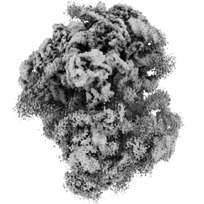 emd_22198.png emd_22198.png | 141.8 KB | ||
| Masks | emd_22198_msk_1.map | 421.9 MB | Mask map | |
| Filedesc metadata | emd-22198.cif.gz | 18.4 KB | ||
| Others | emd_22198_half_map_1.map.gzemd_22198_half_map_2.map.gz | 337.9 MB 338 MB | ||
| Archive directory |  http://ftp.pdbj.org/pub/emdb/structures/EMD-22198ftp://ftp.pdbj.org/pub/emdb/structures/EMD-22198 http://ftp.pdbj.org/pub/emdb/structures/EMD-22198ftp://ftp.pdbj.org/pub/emdb/structures/EMD-22198 | HTTPS FTP |
-Validation report
| Summary document | emd_22198_validation.pdf.gz | 797.3 KB | Display | EMDB validaton report |
|---|---|---|---|---|
| Full document | emd_22198_full_validation.pdf.gz | 796.8 KB | Display | |
| Data in XML | emd_22198_validation.xml.gz | 25.3 KB | Display | |
| Data in CIF | emd_22198_validation.cif.gz | 33.8 KB | Display | |
| Arichive directory | https://ftp.pdbj.org/pub/emdb/validation_reports/EMD-22198ftp://ftp.pdbj.org/pub/emdb/validation_reports/EMD-22198 | HTTPS FTP |
-Related structure data
| Related structure data |  6xirMC  6xiqC C: citing same article ( M: atomic model generated by this map |
|---|---|
| Similar structure data |

-Links
| EMDB pages | EMDB (EBI/PDBe) / EMDataResource |
|---|---|
| Related items in Molecule of the Month |


-Map
| File | Download / File: emd_22198.map.gz / Format: CCP4 / Size: 421.9 MB / Type: IMAGE STORED AS FLOATING POINT NUMBER (4 BYTES) | ||||||||||||||||||||||||||||||||||||||||||||||||||||||||||||||||||||
|---|---|---|---|---|---|---|---|---|---|---|---|---|---|---|---|---|---|---|---|---|---|---|---|---|---|---|---|---|---|---|---|---|---|---|---|---|---|---|---|---|---|---|---|---|---|---|---|---|---|---|---|---|---|---|---|---|---|---|---|---|---|---|---|---|---|---|---|---|---|
| Voxel size | X=Y=Z: 1.36 Å | ||||||||||||||||||||||||||||||||||||||||||||||||||||||||||||||||||||
| Density |
| ||||||||||||||||||||||||||||||||||||||||||||||||||||||||||||||||||||
| Symmetry | Space group: 1 | ||||||||||||||||||||||||||||||||||||||||||||||||||||||||||||||||||||
| Details | EMDB XML:
CCP4 map header:
| ||||||||||||||||||||||||||||||||||||||||||||||||||||||||||||||||||||
-Supplemental data
- Sample components
+Entire : K63 ubiquitinated Ribosome
+Supramolecule #1: K63 ubiquitinated Ribosome
+Macromolecule #1: 60S ribosomal protein L2-A
+Macromolecule #2: RPL3 isoform 1
+Macromolecule #3: RPL4A isoform 1
+Macromolecule #4: RPL5 isoform 1
+Macromolecule #5: 60S ribosomal protein L6-A
+Macromolecule #6: 60S ribosomal protein L7-A
+Macromolecule #7: RPL8A isoform 1
+Macromolecule #8: RPL9A isoform 1
+Macromolecule #9: RPL10 isoform 1
+Macromolecule #10: RPL11B isoform 1
+Macromolecule #11: 60S ribosomal protein L13-A
+Macromolecule #12: 60S ribosomal protein L14-A
+Macromolecule #13: 60S ribosomal protein L15-A
+Macromolecule #14: 60S ribosomal protein L16-A
+Macromolecule #15: 60S ribosomal protein L17-A
+Macromolecule #16: 60S ribosomal protein L18-A
+Macromolecule #17: 60S ribosomal protein L19-A
+Macromolecule #18: 60S ribosomal protein L20-A
+Macromolecule #19: 60S ribosomal protein L21-A
+Macromolecule #20: 60S ribosomal protein L22-A
+Macromolecule #21: 60S ribosomal protein L23-A
+Macromolecule #22: RPL24A isoform 1
+Macromolecule #23: 60S ribosomal protein L25
+Macromolecule #24: 60S ribosomal protein L26-A
+Macromolecule #25: 60S ribosomal protein L27-A
+Macromolecule #26: 40S ribosomal protein S10-A
+Macromolecule #27: 40S ribosomal protein S11-A
+Macromolecule #31: 60S ribosomal protein L28
+Macromolecule #32: RPL29 isoform 1
+Macromolecule #33: 60S ribosomal protein L30
+Macromolecule #34: 60S ribosomal protein L31-A
+Macromolecule #35: RPL32 isoform 1
+Macromolecule #36: 60S ribosomal protein L33-A
+Macromolecule #37: 60S ribosomal protein L34-A
+Macromolecule #38: 60S ribosomal protein L35-A
+Macromolecule #39: 60S ribosomal protein L36-A
+Macromolecule #40: 60S ribosomal protein L37-A
+Macromolecule #41: RPL38 isoform 1
+Macromolecule #42: 60S ribosomal protein L39
+Macromolecule #43: RPL41A isoform 1
+Macromolecule #44: 60S ribosomal protein L42-A
+Macromolecule #45: 60S ribosomal protein L43-A
+Macromolecule #47: 40S ribosomal protein S0-A
+Macromolecule #48: RPS1A isoform 1
+Macromolecule #49: RPS2 isoform 1
+Macromolecule #50: RPS3 isoform 1
+Macromolecule #51: 40S ribosomal protein S4-A
+Macromolecule #52: Rps5p
+Macromolecule #53: 40S ribosomal protein S6-A
+Macromolecule #54: 40S ribosomal protein S7-A
+Macromolecule #55: RPS8A isoform 1
+Macromolecule #56: 40S ribosomal protein S9-A
+Macromolecule #57: 40S ribosomal protein S13
+Macromolecule #58: 40S ribosomal protein S14-B
+Macromolecule #59: RPS15 isoform 1
+Macromolecule #60: 40S ribosomal protein S16-A
+Macromolecule #61: 40S ribosomal protein S17-B
+Macromolecule #62: 40S ribosomal protein S18-A
+Macromolecule #63: 40S ribosomal protein S19-A
+Macromolecule #64: RPS20 isoform 1
+Macromolecule #65: 40S ribosomal protein S21-A
+Macromolecule #66: RPS22A isoform 1
+Macromolecule #67: 40S ribosomal protein S23-A
+Macromolecule #68: 40S ribosomal protein S24-A
+Macromolecule #69: RPS25A isoform 1
+Macromolecule #70: RPS26B isoform 1
+Macromolecule #71: 40S ribosomal protein S27-A
+Macromolecule #72: RPS28A isoform 1
+Macromolecule #73: RPS29A isoform 1
+Macromolecule #74: Guanine nucleotide-binding protein subunit beta-like protein
+Macromolecule #28: 35S ribosomal RNA
+Macromolecule #29: 5S ribosomal RNA
+Macromolecule #30: 5.8S ribosomal RNA
+Macromolecule #46: 18S ribosomal RNA
+Macromolecule #75: Transfer RNA
+Macromolecule #76: ZINC ION
-Experimental details
-Structure determination
| Method | cryo EM |
|---|---|
 Processing Processing | single particle reconstruction |
| Aggregation state | particle |
-Sample preparation
| Buffer | pH: 7.5 |
|---|---|
| Grid | Model: Quantifoil R2/1 / Material: COPPER / Support film - Material: CARBON / Support film - topology: HOLEY |
| Vitrification | Cryogen name: ETHANE |
- Electron microscopy
| Microscope | FEI TITAN KRIOS |
|---|---|
| Image recording | Film or detector model: FEI FALCON II (4k x 4k) / Detector mode: INTEGRATING / Digitization - Frames/image: 1-7 / Number real images: 5243 / Average electron dose: 25.0 e/Å2 |
| Electron beam | Acceleration voltage: 300 kV / Electron source:  FIELD EMISSION GUN FIELD EMISSION GUN |
| Electron optics | Illumination mode: FLOOD BEAM / Imaging mode: BRIGHT FIELD |
| Sample stage | Specimen holder model: FEI TITAN KRIOS AUTOGRID HOLDER / Cooling holder cryogen: NITROGEN |
| Experimental equipment |  Model: Titan Krios / Image courtesy: FEI Company |
-Image processing
| Startup model | Type of model: INSILICO MODEL |
|---|---|
| Final reconstruction | Number classes used: 1 / Applied symmetry - Point group: C1 (asymmetric) / Algorithm: FOURIER SPACE / Resolution.type: BY AUTHOR / Resolution: 3.2 Å / Resolution method: FSC 0.143 CUT-OFF / Software - Name: RELION (ver. 3.0) / Number images used: 87939 |
| Initial angle assignment | Type: MAXIMUM LIKELIHOOD / Software - Name: cryoSPARC (ver. 2.12.0) |
| Final angle assignment | Type: MAXIMUM LIKELIHOOD / Software - Name: RELION (ver. 3.0) |