 ムービー
ムービー コントローラー


+ データを開く

- 基本情報
基本情報
| 登録情報 | データベース: EMDB / ID: EMD-21706 | |||||||||||||||
|---|---|---|---|---|---|---|---|---|---|---|---|---|---|---|---|---|
| タイトル | Cryogenic Single-Molecule Fluorescence Annotations for Electron Tomography Reveal In Situ Organization of Key Proteins in Caulobacter | |||||||||||||||
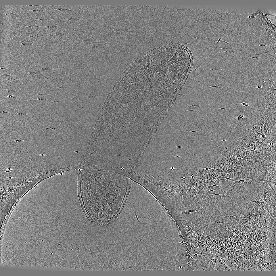
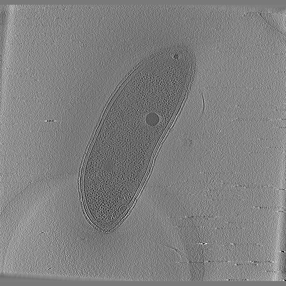
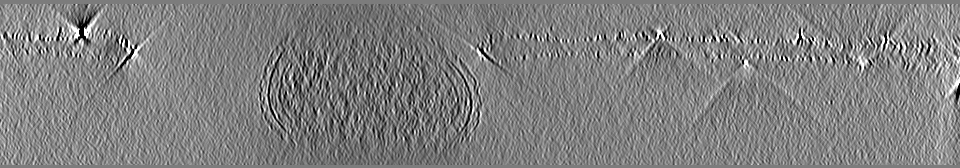
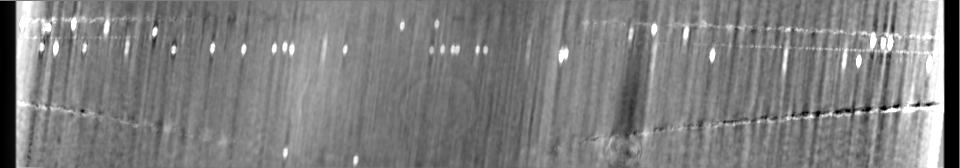
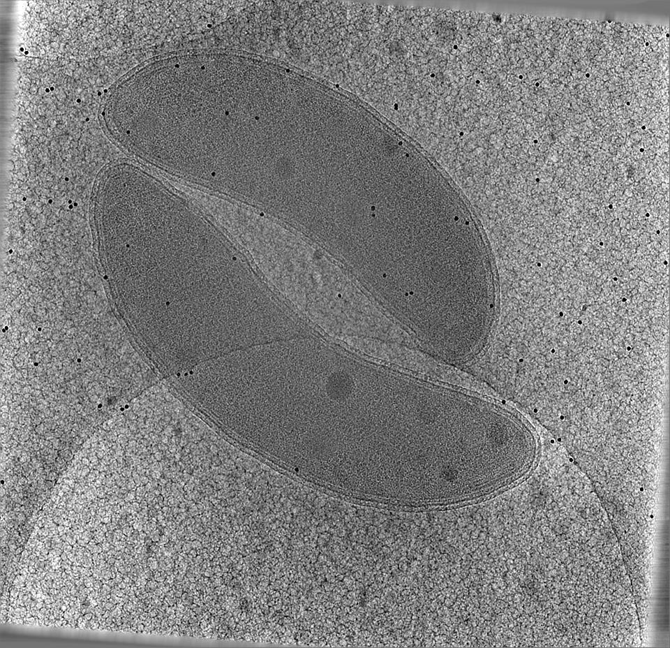
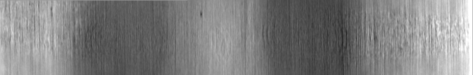
 マップデータ マップデータ | Tomographic Reconstruction (bin by 4) of C. Crescentus that underwent no correlative imaging. | |||||||||||||||
 試料 試料 |
| |||||||||||||||
 キーワード キーワード | Bacteria / Tomography / Regulatory proteins / CELL CYCLE | |||||||||||||||
| 生物種 |  Caulobacter vibrioides (バクテリア) Caulobacter vibrioides (バクテリア) | |||||||||||||||
| 手法 | 電子線トモグラフィー法 / クライオ電子顕微鏡法 | |||||||||||||||
 データ登録者 データ登録者 | Dahlberg PD / Saurabh S / Sartor AM / Wang J / Mitchell P / Chiu W / Shapiro L / Moerner WE | |||||||||||||||
| 資金援助 |  米国, 4件 米国, 4件
| |||||||||||||||
 引用 引用 | ジャーナル: Proc Natl Acad Sci U S A / 年: 2020 タイトル: Cryogenic single-molecule fluorescence annotations for electron tomography reveal in situ organization of key proteins in . 著者: Peter D Dahlberg / Saumya Saurabh / Annina M Sartor / Jiarui Wang / Patrick G Mitchell / Wah Chiu / Lucy Shapiro / W E Moerner / 要旨: Superresolution fluorescence microscopy and cryogenic electron tomography (CET) are powerful imaging methods for exploring the subcellular organization of biomolecules. Superresolution fluorescence ...Superresolution fluorescence microscopy and cryogenic electron tomography (CET) are powerful imaging methods for exploring the subcellular organization of biomolecules. Superresolution fluorescence microscopy based on covalent labeling highlights specific proteins and has sufficient sensitivity to observe single fluorescent molecules, but the reconstructions lack detailed cellular context. CET has molecular-scale resolution but lacks specific and nonperturbative intracellular labeling techniques. Here, we describe an imaging scheme that correlates cryogenic single-molecule fluorescence localizations with CET reconstructions. Our approach achieves single-molecule localizations with an average lateral precision of 9 nm, and a relative registration error between the set of localizations and CET reconstruction of ∼30 nm. We illustrate the workflow by annotating the positions of three proteins in the bacterium : McpA, PopZ, and SpmX. McpA, which forms a part of the chemoreceptor array, acts as a validation structure by being visible under both imaging modalities. In contrast, PopZ and SpmX cannot be directly identified in CET. While not directly discernable, PopZ fills a region at the cell poles that is devoid of electron-dense ribosomes. We annotate the position of PopZ with single-molecule localizations and confirm its position within the ribosome excluded region. We further use the locations of PopZ to provide context for localizations of SpmX, a low-copy integral membrane protein sequestered by PopZ as part of a signaling pathway that leads to an asymmetric cell division. Our correlative approach reveals that SpmX localizes along one side of the cell pole and its extent closely matches that of the PopZ region. | |||||||||||||||
| 履歴 |
|
- 構造の表示
構造の表示
| ムービー |
 ムービービューア ムービービューア |
|---|---|
| 添付画像 |

- ダウンロードとリンク
-EMDBアーカイブ
| マップデータ | emd_21706.map.gz | 130.1 MB | EMDBマップデータ形式 | |
|---|---|---|---|---|
| ヘッダ (付随情報) | emd-21706-v30.xmlemd-21706.xml | 22 KB 22 KB | 表示 表示 | EMDBヘッダ |
| 画像 | 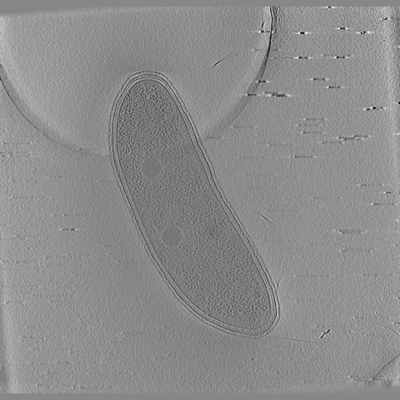 emd_21706.png emd_21706.png | 97.6 KB | ||
| その他 | emd_21706_additional_1.map.gzemd_21706_additional_2.map.gzemd_21706_additional_3.map.gzemd_21706_additional_4.map.gzemd_21706_additional_5.map.gz | 543.6 MB 589.7 KB 178.8 MB 774.5 KB 118.2 MB | ||
| アーカイブディレクトリ |  http://ftp.pdbj.org/pub/emdb/structures/EMD-21706ftp://ftp.pdbj.org/pub/emdb/structures/EMD-21706 http://ftp.pdbj.org/pub/emdb/structures/EMD-21706ftp://ftp.pdbj.org/pub/emdb/structures/EMD-21706 | HTTPS FTP |
-検証レポート
| 文書・要旨 | emd_21706_validation.pdf.gz | 358.6 KB | 表示 | EMDB検証レポート |
|---|---|---|---|---|
| 文書・詳細版 | emd_21706_full_validation.pdf.gz | 358.2 KB | 表示 | |
| XML形式データ | emd_21706_validation.xml.gz | 4.8 KB | 表示 | |
| CIF形式データ | emd_21706_validation.cif.gz | 5.3 KB | 表示 | |
| アーカイブディレクトリ | https://ftp.pdbj.org/pub/emdb/validation_reports/EMD-21706ftp://ftp.pdbj.org/pub/emdb/validation_reports/EMD-21706 | HTTPS FTP |
-リンク
| EMDBのページ | EMDB (EBI/PDBe) / EMDataResource |
|---|
-マップ
| ファイル | ダウンロード / ファイル: emd_21706.map.gz / 形式: CCP4 / 大きさ: 590.6 MB / タイプ: IMAGE STORED AS FLOATING POINT NUMBER (4 BYTES) | ||||||||||||||||||||||||||||||||||||||||||||||||||||||||||||||||||||
|---|---|---|---|---|---|---|---|---|---|---|---|---|---|---|---|---|---|---|---|---|---|---|---|---|---|---|---|---|---|---|---|---|---|---|---|---|---|---|---|---|---|---|---|---|---|---|---|---|---|---|---|---|---|---|---|---|---|---|---|---|---|---|---|---|---|---|---|---|---|
| 注釈 | Tomographic Reconstruction (bin by 4) of C. Crescentus that underwent no correlative imaging. | ||||||||||||||||||||||||||||||||||||||||||||||||||||||||||||||||||||
| 投影像・断面図 | 画像のコントロール
画像は Spider により作成 これらの図は立方格子座標系で作成されたものです | ||||||||||||||||||||||||||||||||||||||||||||||||||||||||||||||||||||
| ボクセルのサイズ | X=Y=Z: 29.16 Å | ||||||||||||||||||||||||||||||||||||||||||||||||||||||||||||||||||||
| 密度 |
| ||||||||||||||||||||||||||||||||||||||||||||||||||||||||||||||||||||
| 対称性 | 空間群: 1 | ||||||||||||||||||||||||||||||||||||||||||||||||||||||||||||||||||||
| 詳細 | EMDB XML:
CCP4マップ ヘッダ情報:
| ||||||||||||||||||||||||||||||||||||||||||||||||||||||||||||||||||||
 Z (Sec.)
 Y (Row.)
 X (Col.)


-添付データ
-追加マップ: Tomographic Reconstruction (bin 4) of C. Crescentus that...
| ファイル | emd_21706_additional_1.map | ||||||||||||
|---|---|---|---|---|---|---|---|---|---|---|---|---|---|
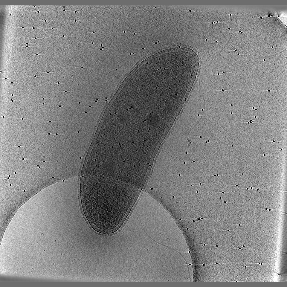
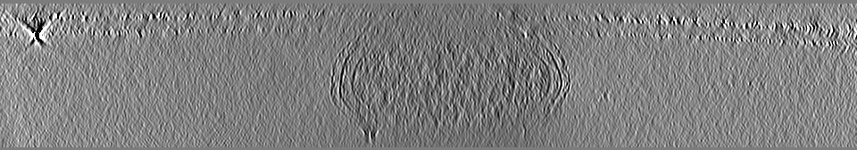
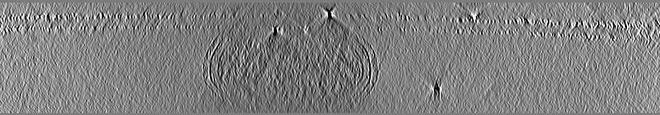
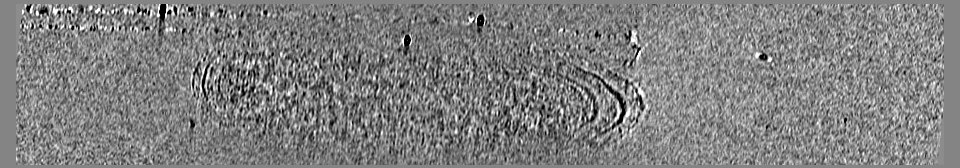
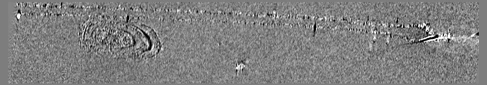
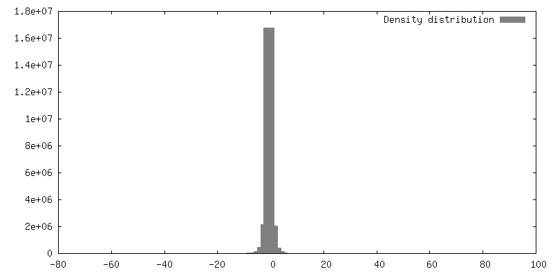
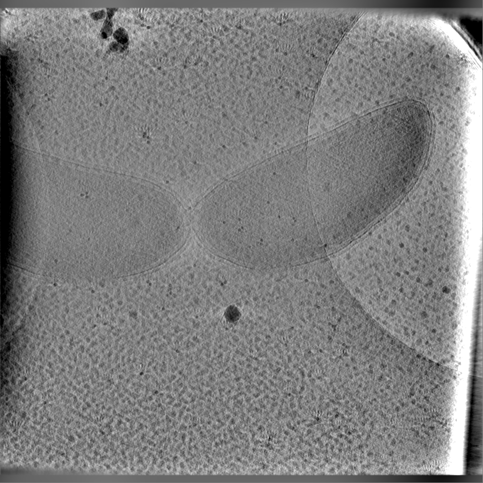
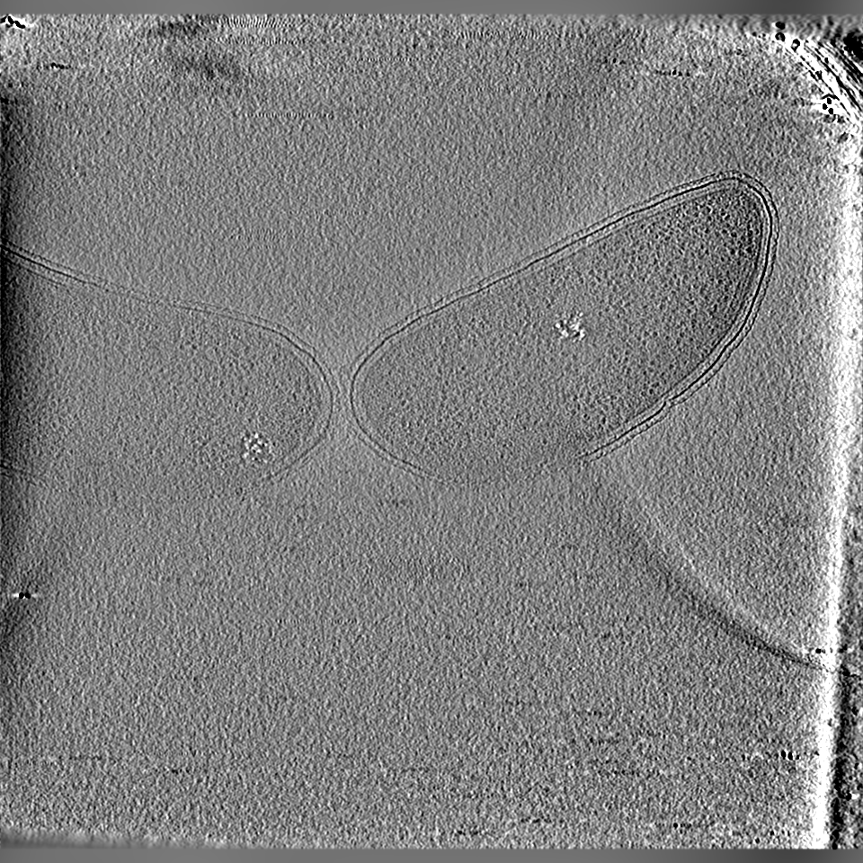
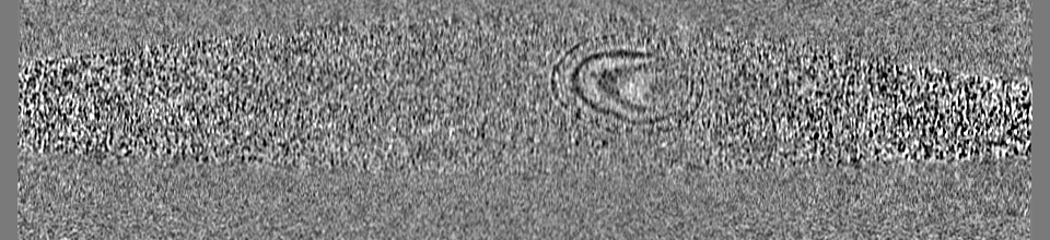
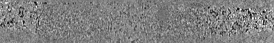
| 注釈 | Tomographic Reconstruction (bin 4) of C. Crescentus that underwent correlative imaging. See corresponding file for single-molecule fluorescence localizations annotating the positions of PAmKate-PopZ. | ||||||||||||
| 投影像・断面図 |
| ||||||||||||
| 密度ヒストグラム |


-追加マップ: Single-molecule fluorescence localizations annotating the positions of PAmKate-PopZ....
| ファイル | emd_21706_additional_2.map | ||||||||||||
|---|---|---|---|---|---|---|---|---|---|---|---|---|---|
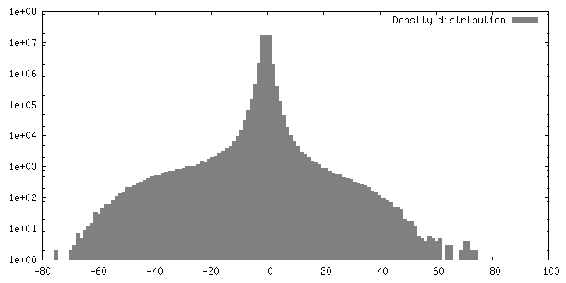
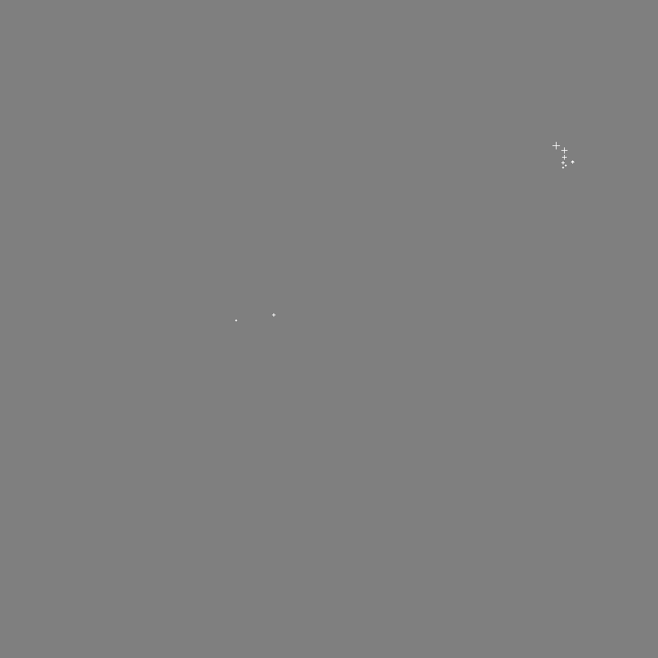
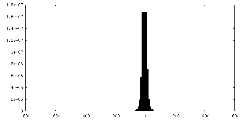
| 注釈 | Single-molecule fluorescence localizations annotating the positions of PAmKate-PopZ. | ||||||||||||
| 投影像・断面図 |
| ||||||||||||
| 密度ヒストグラム |


-追加マップ: Tomographic Reconstruction (bin 4) of C. Crescentus that...
| ファイル | emd_21706_additional_3.map | ||||||||||||
|---|---|---|---|---|---|---|---|---|---|---|---|---|---|
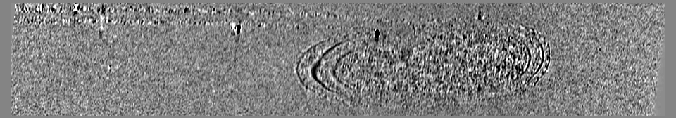
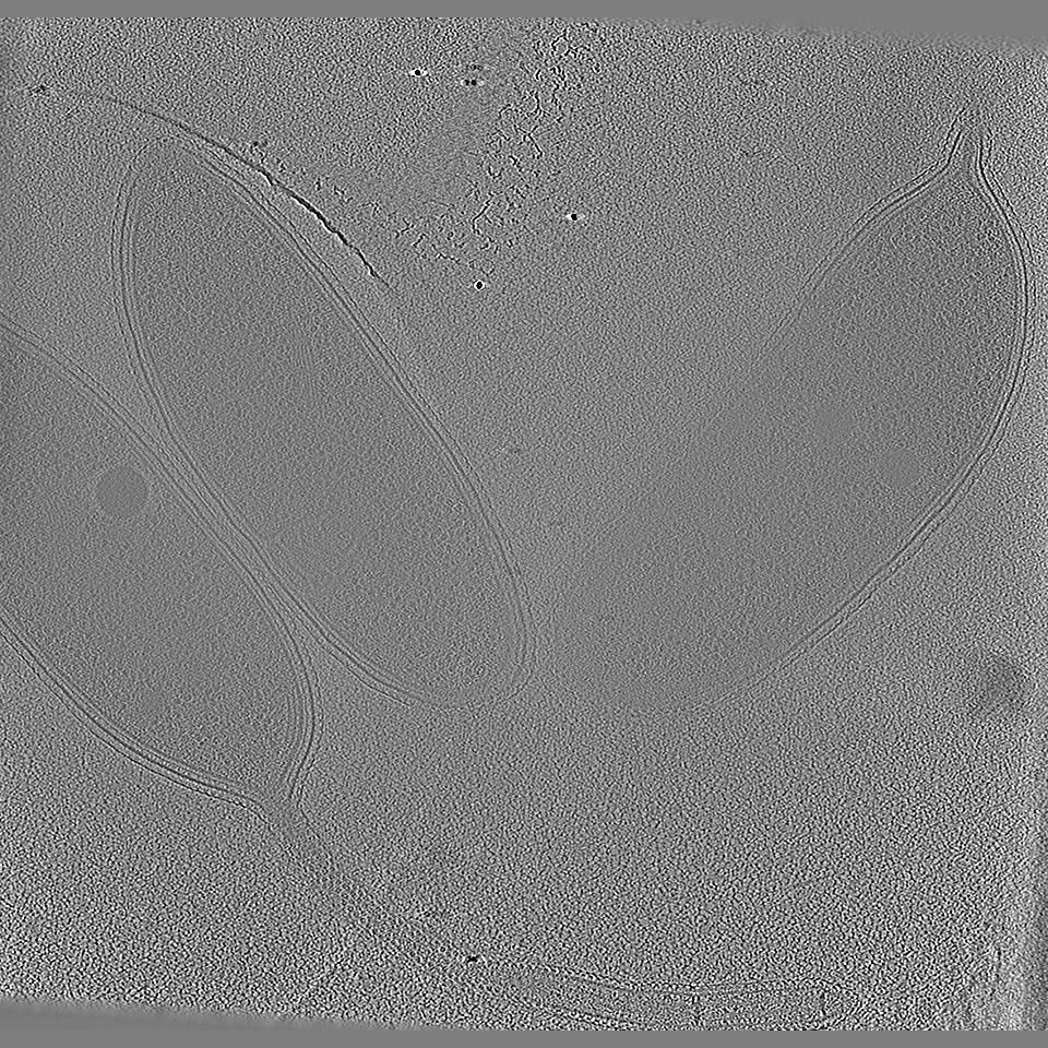
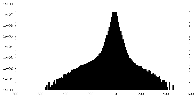
| 注釈 | Tomographic Reconstruction (bin 4) of C. Crescentus that underwent correlative imaging. See corresponding file for single-molecule fluorescence localizations annotating the positions of PAmKate-McpA. | ||||||||||||
| 投影像・断面図 |
| ||||||||||||
| 密度ヒストグラム |


-追加マップ: Single-molecule fluorescence localizations annotating the positions of PAmKate-McpA....
| ファイル | emd_21706_additional_4.map | ||||||||||||
|---|---|---|---|---|---|---|---|---|---|---|---|---|---|
| 注釈 | Single-molecule fluorescence localizations annotating the positions of PAmKate-McpA. | ||||||||||||
| 投影像・断面図 |
| ||||||||||||
| 密度ヒストグラム |


-追加マップ: Tomographic Reconstruction (bin by 4) of C. Crescentus...
| ファイル | emd_21706_additional_5.map | ||||||||||||
|---|---|---|---|---|---|---|---|---|---|---|---|---|---|
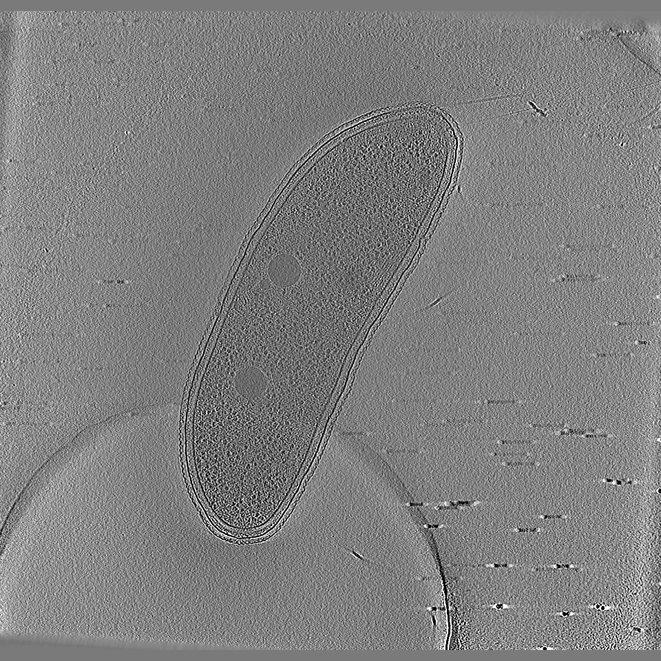
| 注釈 | Tomographic Reconstruction (bin by 4) of C. Crescentus that underwent correlative imaging. Fluorescence imaging was performed using an excitation beam with an intensity of ~80W/cm^2 for ~3 hours. | ||||||||||||
| 投影像・断面図 |
| ||||||||||||
| 密度ヒストグラム |


- 試料の構成要素
-全体 : Caulobacter crescentus cells
| 全体 | 名称: Caulobacter crescentus cells |
|---|---|
| 要素 |
|
-超分子 #1: Caulobacter crescentus cells
| 超分子 | 名称: Caulobacter crescentus cells / タイプ: cell / ID: 1 / 親要素: 0 |
|---|---|
| 由来(天然) | 生物種: Caulobacter vibrioides (バクテリア) |
-実験情報
-構造解析
| 手法 | クライオ電子顕微鏡法 |
|---|---|
 解析 解析 | 電子線トモグラフィー法 |
| 試料の集合状態 | cell |
-試料調製
| 緩衝液 | pH: 6.8 / 詳細: Cells cultured and vitrified in M2G growth media |
|---|---|
| グリッド | モデル: Quantifoil R2/2 / 材質: COPPER / メッシュ: 200 / 支持フィルム - 材質: CARBON / 支持フィルム - トポロジー: HOLEY / 前処理 - タイプ: GLOW DISCHARGE |
| 凍結 | 凍結剤: ETHANE / チャンバー内湿度: 95 % / チャンバー内温度: 298 K / 装置: GATAN CRYOPLUNGE 3 / 詳細: double sided blot for 3.5 seconds. |
| Cryo protectant | 20% ethylene glycol |
| 切片作成 | その他: NO SECTIONING |
| 位置合わせマーカー | Manufacturer: EMS / 直径: 15 nm |
- 電子顕微鏡法
| 顕微鏡 | TFS KRIOS |
|---|---|
| 特殊光学系 | エネルギーフィルター - 名称: GIF Bioquantum / エネルギーフィルター - スリット幅: 20 eV |
| 撮影 | フィルム・検出器のモデル: GATAN K2 SUMMIT (4k x 4k) 検出モード: COUNTING / デジタル化 - サイズ - 横: 3838 pixel / デジタル化 - サイズ - 縦: 3710 pixel / 平均電子線量: 1.1 e/Å2 |
| 電子線 | 加速電圧: 300 kV / 電子線源:  FIELD EMISSION GUN FIELD EMISSION GUN |
| 電子光学系 | 照射モード: FLOOD BEAM / 撮影モード: BRIGHT FIELD / Cs: 2.7 mm / 最小 デフォーカス(公称値): 10.0 µm |
| 試料ステージ | 試料ホルダーモデル: FEI TITAN KRIOS AUTOGRID HOLDER ホルダー冷却材: NITROGEN |
| 実験機器 |  モデル: Titan Krios / 画像提供: FEI Company |
-画像解析
| 最終 再構成 | アルゴリズム: BACK PROJECTION / ソフトウェア - 名称: IMOD / 使用した粒子像数: 91 |
|---|
