 Movie
Movie Controller

[English] 日本語
 Yorodumi
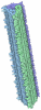
- EMDB-21501: Amyloid-beta(1-40) fibril derived from Alzheimer's disease cortic... -
+ Open data

- Basic information
| Entry | Database: EMDB / ID: EMD-21501 | |||||||||
|---|---|---|---|---|---|---|---|---|---|---|
| Title | Amyloid-beta(1-40) fibril derived from Alzheimer's disease cortical tissue | |||||||||
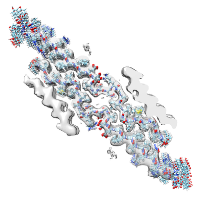
 Map data Map data | CryoEM density map from RELION-based analysis | |||||||||
 Sample Sample |
| |||||||||
 Keywords Keywords | amyloid-beta / Alzheimer's disease / PROTEIN FIBRIL | |||||||||
| Function / homology |  Function and homology information Function and homology informationamyloid-beta complex / growth cone lamellipodium / cellular response to norepinephrine stimulus / collateral sprouting in absence of injury / growth cone filopodium / microglia development / hippocampal neuron apoptotic process / regulation of Wnt signaling pathway / Formyl peptide receptors bind formyl peptides and many other ligands / axo-dendritic transport ...amyloid-beta complex / growth cone lamellipodium / cellular response to norepinephrine stimulus / collateral sprouting in absence of injury / growth cone filopodium / microglia development / hippocampal neuron apoptotic process / regulation of Wnt signaling pathway / Formyl peptide receptors bind formyl peptides and many other ligands / axo-dendritic transport / axon midline choice point recognition / regulation of synapse structure or activity / positive regulation of synaptic transmission, cholinergic / astrocyte activation involved in immune response / NMDA selective glutamate receptor signaling pathway / regulation of spontaneous synaptic transmission / mating behavior / growth factor receptor binding / Insertion of tail-anchored proteins into the endoplasmic reticulum membrane / positive regulation of amyloid fibril formation / peptidase activator activity / PTB domain binding / Golgi-associated vesicle / Lysosome Vesicle Biogenesis / Deregulated CDK5 triggers multiple neurodegenerative pathways in Alzheimer's disease models / neuron remodeling / astrocyte projection / regulation of multicellular organism growth / nuclear envelope lumen / dendrite development / TRAF6 mediated NF-kB activation / positive regulation of protein metabolic process / negative regulation of long-term synaptic potentiation / signaling receptor activator activity / Notch signaling pathway / transition metal ion binding / Advanced glycosylation endproduct receptor signaling / The NLRP3 inflammasome / intracellular copper ion homeostasis / modulation of excitatory postsynaptic potential / main axon / ECM proteoglycans / positive regulation of T cell migration / response to insulin-like growth factor stimulus / regulation of presynapse assembly / extracellular matrix organization / swimming behavior / adult locomotory behavior / neuronal dense core vesicle / regulation of long-term neuronal synaptic plasticity / Purinergic signaling in leishmaniasis infection / positive regulation of calcium-mediated signaling / positive regulation of chemokine production / positive regulation of mitotic cell cycle / axonogenesis / cellular response to manganese ion / neuron projection maintenance / clathrin-coated pit / visual learning / cellular response to cAMP / astrocyte activation / synaptic cleft / Mitochondrial protein degradation / response to interleukin-1 / platelet alpha granule lumen / regulation of neuron apoptotic process / positive regulation of glycolytic process / learning / ionotropic glutamate receptor signaling pathway / locomotory behavior / cellular response to copper ion / endosome lumen / positive regulation of interleukin-1 beta production / central nervous system development / positive regulation of long-term synaptic potentiation / protein serine/threonine kinase binding / serine-type endopeptidase inhibitor activity / Post-translational protein phosphorylation / dendritic shaft / trans-Golgi network membrane / endocytosis / microglial cell activation / cellular response to nerve growth factor stimulus / regulation of translation / positive regulation of non-canonical NF-kappaB signal transduction / positive regulation of interleukin-6 production / positive regulation of JNK cascade / synapse organization / TAK1-dependent IKK and NF-kappa-B activation / Golgi lumen / recycling endosome / response to lead ion / cognition / Regulation of Insulin-like Growth Factor (IGF) transport and uptake by Insulin-like Growth Factor Binding Proteins (IGFBPs) / cellular response to amyloid-beta / neuron projection development / calcium ion transport / positive regulation of inflammatory response / regulation of gene expression / positive regulation of tumor necrosis factor production Similarity search - Function | |||||||||
| Biological species |  Homo sapiens (human) Homo sapiens (human) | |||||||||
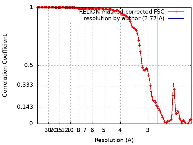
| Method | helical reconstruction / cryo EM / Resolution: 2.77 Å | |||||||||
 Authors Authors | Ghosh U / Thurber KR | |||||||||
 Citation Citation | Journal: Proc Natl Acad Sci U S A / Year: 2021 Title: Molecular structure of a prevalent amyloid-β fibril polymorph from Alzheimer's disease brain tissue. Authors: Ujjayini Ghosh / Kent R Thurber / Wai-Ming Yau / Robert Tycko /  Abstract: Amyloid-β (Aβ) fibrils exhibit self-propagating, molecular-level polymorphisms that may contribute to variations in clinical and pathological characteristics of Alzheimer's disease (AD). We report ...Amyloid-β (Aβ) fibrils exhibit self-propagating, molecular-level polymorphisms that may contribute to variations in clinical and pathological characteristics of Alzheimer's disease (AD). We report the molecular structure of a specific fibril polymorph, formed by 40-residue Aβ peptides (Aβ40), that is derived from cortical tissue of an AD patient by seeded fibril growth. The structure is determined from cryogenic electron microscopy (cryoEM) images, supplemented by mass-per-length (MPL) measurements and solid-state NMR (ssNMR) data. Previous ssNMR studies with multiple AD patients had identified this polymorph as the most prevalent brain-derived Aβ40 fibril polymorph from typical AD patients. The structure, which has 2.8-Å resolution according to standard criteria, differs qualitatively from all previously described Aβ fibril structures, both in its molecular conformations and its organization of cross-β subunits. Unique features include twofold screw symmetry about the fibril growth axis, despite an MPL value that indicates three Aβ40 molecules per 4.8-Å β-sheet spacing, a four-layered architecture, and fully extended conformations for molecules in the central two cross-β layers. The cryoEM density, ssNMR data, and MPL data are consistent with β-hairpin conformations for molecules in the outer cross-β layers. Knowledge of this brain-derived fibril structure may contribute to the development of structure-specific amyloid imaging agents and aggregation inhibitors with greater diagnostic and therapeutic utility. | |||||||||
| History |
|
- Structure visualization
| Movie |
Movie viewer |
|---|---|
| Structure viewer | EM map: SurfViewMolmilJmol/JSmol |
| Supplemental images |
- Downloads & links
-EMDB archive
| Map data | emd_21501.map.gz | 224.1 MB | EMDB map data format | |
|---|---|---|---|---|
| Header (meta data) | emd-21501-v30.xmlemd-21501.xml | 14.3 KB 14.3 KB | Display Display | EMDB header |
| FSC (resolution estimation) | emd_21501_fsc.xml | 14.1 KB | Display | FSC data file |
| Images |  emd_21501.png emd_21501.png | 120.4 KB | ||
| Masks | emd_21501_msk_1.map | 244.1 MB | Mask map | |
| Filedesc metadata | emd-21501.cif.gz | 6.1 KB | ||
| Archive directory |  https://data.pdbj.org/pub/emdb/structures/EMD-21501ftp://data.pdbj.org/pub/emdb/structures/EMD-21501 https://data.pdbj.org/pub/emdb/structures/EMD-21501ftp://data.pdbj.org/pub/emdb/structures/EMD-21501 | HTTPS FTP |
-Related structure data
| Related structure data |  6w0oMC M: atomic model generated by this map C: citing same article ( |
|---|---|
| Similar structure data |

-Links
| EMDB pages | EMDB (EBI/PDBe) / EMDataResource |
|---|---|
| Related items in Molecule of the Month |




-Map
| File | Download / File: emd_21501.map.gz / Format: CCP4 / Size: 244.1 MB / Type: IMAGE STORED AS FLOATING POINT NUMBER (4 BYTES) | ||||||||||||||||||||||||||||||||||||||||||||||||||||||||||||||||||||
|---|---|---|---|---|---|---|---|---|---|---|---|---|---|---|---|---|---|---|---|---|---|---|---|---|---|---|---|---|---|---|---|---|---|---|---|---|---|---|---|---|---|---|---|---|---|---|---|---|---|---|---|---|---|---|---|---|---|---|---|---|---|---|---|---|---|---|---|---|---|
| Annotation | CryoEM density map from RELION-based analysis | ||||||||||||||||||||||||||||||||||||||||||||||||||||||||||||||||||||
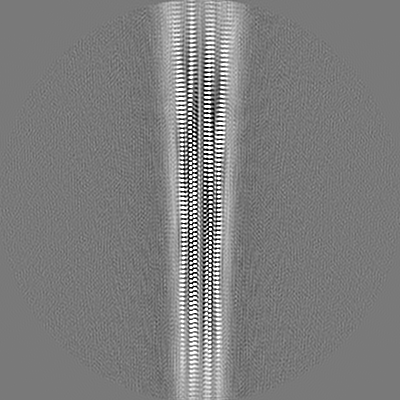
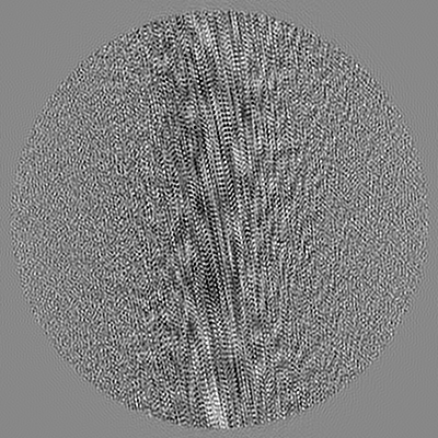
| Projections & slices | Image control
Images are generated by Spider. | ||||||||||||||||||||||||||||||||||||||||||||||||||||||||||||||||||||
| Voxel size | X=Y=Z: 1.08 Å | ||||||||||||||||||||||||||||||||||||||||||||||||||||||||||||||||||||
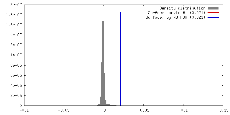
| Density |
| ||||||||||||||||||||||||||||||||||||||||||||||||||||||||||||||||||||
| Symmetry | Space group: 1 | ||||||||||||||||||||||||||||||||||||||||||||||||||||||||||||||||||||
| Details | EMDB XML:
CCP4 map header:
| ||||||||||||||||||||||||||||||||||||||||||||||||||||||||||||||||||||
 Z (Sec.)
 Y (Row.)
 X (Col.)






-Supplemental data
-Mask #1
| File | emd_21501_msk_1.map | ||||||||||||
|---|---|---|---|---|---|---|---|---|---|---|---|---|---|
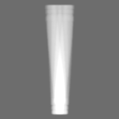
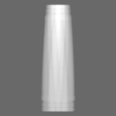
| Projections & Slices |
| ||||||||||||
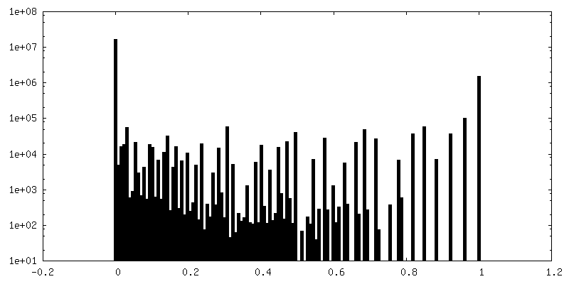
| Density Histograms |





- Sample components
-Entire : amyloid-beta(1-40) fibrils derived from human AD brain
| Entire | Name: amyloid-beta(1-40) fibrils derived from human AD brain |
|---|---|
| Components |
|
-Supramolecule #1: amyloid-beta(1-40) fibrils derived from human AD brain
| Supramolecule | Name: amyloid-beta(1-40) fibrils derived from human AD brain type: complex / ID: 1 / Parent: 0 / Macromolecule list: all Details: Fibrils produced by seeded growth using amyloid-beta in brain extract as the source of seeds. CryoEM and solid state NMR measurements were performed on second-generation seeded fibrils. |
|---|---|
| Source (natural) | Organism: Homo sapiens (human) |
| Molecular weight | Theoretical: 29 kDa/nm |
-Macromolecule #1: Amyloid-beta precursor protein
| Macromolecule | Name: Amyloid-beta precursor protein / type: protein_or_peptide / ID: 1 / Number of copies: 6 / Enantiomer: LEVO |
|---|---|
| Source (natural) | Organism: Homo sapiens (human) |
| Molecular weight | Theoretical: 4.335852 KDa |
| Sequence | String: DAEFRHDSGY EVHHQKLVFF AEDVGSNKGA IIGLMVGGVV UniProtKB: Amyloid-beta precursor protein |
-Experimental details
-Structure determination
| Method | cryo EM |
|---|---|
 Processing Processing | helical reconstruction |
| Aggregation state | filament |
-Sample preparation
| Concentration | 0.45 mg/mL |
|---|---|
| Buffer | pH: 7.4 / Component - Concentration: 10.0 mM / Component - Formula: Na2HPO4/NaH2PO4 / Component - Name: Phosphate buffer Details: 10 mM phosphate buffer with 0.01% NaN3 to avoid microbial contamination. Buffers were filtered to avoid contamination. |
| Grid | Model: Quantifoil R1.2/1.3 / Material: GOLD / Mesh: 300 / Support film - topology: HOLEY / Pretreatment - Type: GLOW DISCHARGE / Pretreatment - Time: 60 sec. / Pretreatment - Atmosphere: OTHER / Pretreatment - Pressure: 0.036000000000000004 kPa / Details: The grids were checked in microscope prior to use. |
| Vitrification | Cryogen name: ETHANE / Chamber humidity: 90 % / Chamber temperature: 293 K / Instrument: LEICA PLUNGER Details: The grids were preblotted for 10 seconds and blotted for 6 seconds before plunging.. |
| Details | Protein exists in solution as amyloid fibrils of varying lengths. |
- Electron microscopy
| Microscope | TFS KRIOS |
|---|---|
| Alignment procedure | Coma free - Residual tilt: 6.0 mrad |
| Specialist optics | Energy filter - Name: GIF Quantum LS / Energy filter - Slit width: 20 eV |
| Details | Preliminary grid screening was done manually in FEI T12. |
| Image recording | Film or detector model: GATAN K2 QUANTUM (4k x 4k) / Detector mode: SUPER-RESOLUTION / Digitization - Frames/image: 1-50 / Number grids imaged: 1 / Number real images: 1337 / Average exposure time: 10.0 sec. / Average electron dose: 73.5 e/Å2 |
| Electron beam | Acceleration voltage: 300 kV / Electron source:  FIELD EMISSION GUN FIELD EMISSION GUN |
| Electron optics | C2 aperture diameter: 100.0 µm / Illumination mode: FLOOD BEAM / Imaging mode: BRIGHT FIELD / Cs: 2.7 mm / Nominal defocus max: -3.0 µm / Nominal defocus min: -0.5 µm / Nominal magnification: 130000 |
| Sample stage | Specimen holder model: FEI TITAN KRIOS AUTOGRID HOLDER / Cooling holder cryogen: NITROGEN |
| Experimental equipment |  Model: Titan Krios / Image courtesy: FEI Company |
+Image processing
-Atomic model buiding 1
| Details | Xplor-NIH was used to combine EM density with phi/psi restraints from NMR chemical shifts (from Talos-N Version 4.21 Rev 2016.343.11.31). |
|---|---|
| Refinement | Space: REAL / Protocol: AB INITIO MODEL |
| Output model | PDB-6w0o: |
