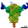
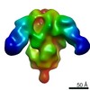
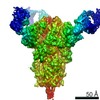
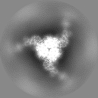
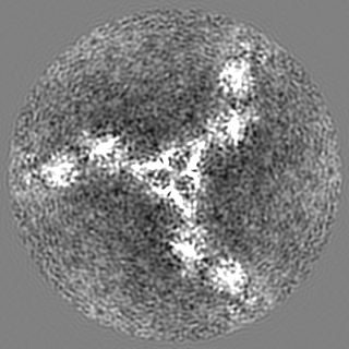
登録情報 データベース : EMDB / ID : EMD-21377タイトル Cryo-electron microscopy structure of mouse coronavirus spike protein complexed with its murine receptor CryoEM density map of MHV spike ectodomain complex with its receptor CEACAM1a 複合体 : MHV Spike and CEACAM1a Complex複合体 : Spike glycoproteinタンパク質・ペプチド : Spike glycoprotein複合体 : Carcinoembryonic antigen-related cell adhesion molecule 1タンパク質・ペプチド : Carcinoembryonic antigen-related cell adhesion molecule 1リガンド : 2-acetamido-2-deoxy-beta-D-glucopyranose / / / / 機能・相同性 分子機能 ドメイン・相同性 構成要素
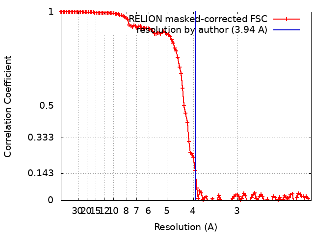
/ / / / / / / / / / / / / / / / / / / / / / / / / / / / / / / / / / / / / / / / / / / / / / / / / / / / / / / / / / / / / / / / / / / / / / / / / / / / / / / / / / / / / / / / / / / / / / / / / / / / / / / / / / / / / / / / / / / / / / / / / / / / / / / / / / / / / / / 生物種 / Mus musculus (ハツカネズミ)手法 / / 解像度 : 3.94 Å Shang J / Wan YS 資金援助 Organization Grant number 国 National Institutes of Health/National Institute Of Allergy and Infectious Diseases (NIH/NIAID) R01AI089728
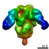
ジャーナル : PLoS Pathog / 年 : 2020タイトル : Structure of mouse coronavirus spike protein complexed with receptor reveals mechanism for viral entry.著者 : Jian Shang / Yushun Wan / Chang Liu / Boyd Yount / Kendra Gully / Yang Yang / Ashley Auerbach / Guiqing Peng / Ralph Baric / Fang Li / 要旨 : Coronaviruses recognize a variety of receptors using different domains of their envelope-anchored spike protein. How these diverse receptor recognition patterns affect viral entry is unknown. Mouse ... Coronaviruses recognize a variety of receptors using different domains of their envelope-anchored spike protein. How these diverse receptor recognition patterns affect viral entry is unknown. Mouse hepatitis coronavirus (MHV) is the only known coronavirus that uses the N-terminal domain (NTD) of its spike to recognize a protein receptor, CEACAM1a. Here we determined the cryo-EM structure of MHV spike complexed with mouse CEACAM1a. The trimeric spike contains three receptor-binding S1 heads sitting on top of a trimeric membrane-fusion S2 stalk. Three receptor molecules bind to the sides of the spike trimer, where three NTDs are located. Receptor binding induces structural changes in the spike, weakening the interactions between S1 and S2. Using protease sensitivity and negative-stain EM analyses, we further showed that after protease treatment of the spike, receptor binding facilitated the dissociation of S1 from S2, allowing S2 to transition from pre-fusion to post-fusion conformation. Together these results reveal a new role of receptor binding in MHV entry: in addition to its well-characterized role in viral attachment to host cells, receptor binding also induces the conformational change of the spike and hence the fusion of viral and host membranes. Our study provides new mechanistic insight into coronavirus entry and highlights the diverse entry mechanisms used by different viruses. 履歴 登録 2020年2月11日 - ヘッダ(付随情報) 公開 2020年3月4日 - マップ公開 2020年3月4日 - 更新 2024年11月6日 - 現状 2024年11月6日 処理サイト : RCSB / 状態 : 公開
すべて表示 表示を減らす
 ムービー
ムービー コントローラー


 データを開く
 基本情報
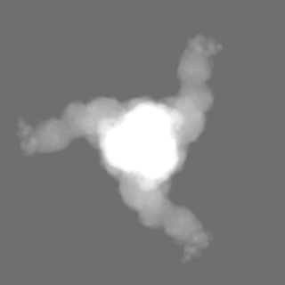
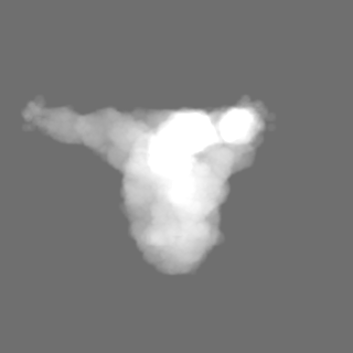
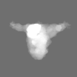
基本情報 マップデータ
 試料
試料 キーワード
キーワード 機能・相同性情報
 Murine coronavirus (strain A59) (ウイルス) /
 データ登録者
 米国, 1件
  引用
引用
 構造の表示
構造の表示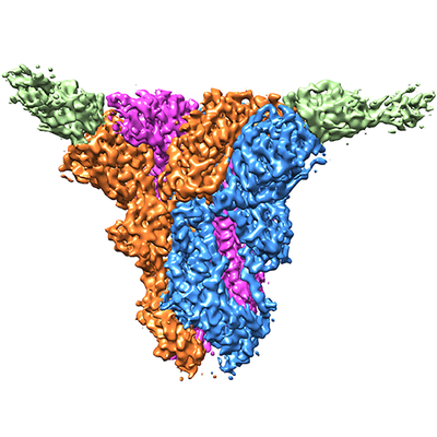
 ダウンロードとリンク
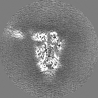
 emd_21377.png
 http://ftp.pdbj.org/pub/emdb/structures/EMD-21377


 Z (Sec.)
 Y (Row.)
 X (Col.)


 試料の構成要素
 解析
解析 電子顕微鏡法
 FIELD EMISSION GUN