ムービー
ムービー コントローラー


+ データを開く

- 基本情報
基本情報
| 登録情報 | データベース: EMDB / ID: EMD-21202 | |||||||||
|---|---|---|---|---|---|---|---|---|---|---|
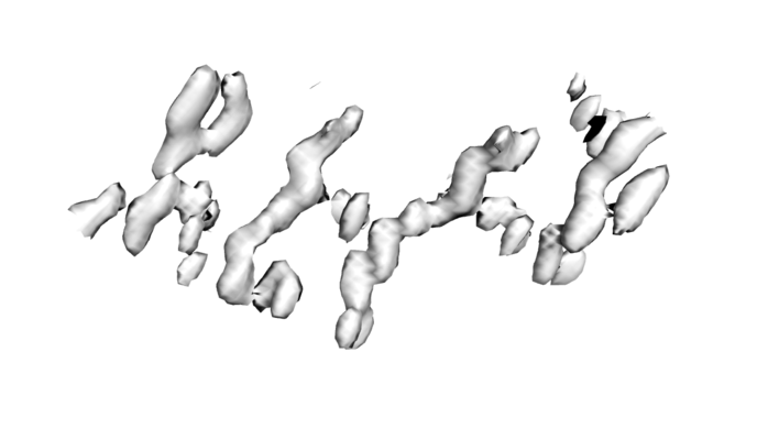
| タイトル | 1.4A low-dose structure of GSNQNNF determined from initial phases generated using radiation damage | |||||||||
 マップデータ マップデータ | 2mFo-dFc map for GSNQNNF built from initial phases generated by radiation damage | |||||||||
 試料 試料 |
| |||||||||
 キーワード キーワード | MicroED / damage / phasing / RIP / protein fibril | |||||||||
| 生物種 | synthetic construct (人工物) | |||||||||
| 手法 | 電子線結晶学 / クライオ電子顕微鏡法 / 解像度: 1.4 Å | |||||||||
 データ登録者 データ登録者 | Martynowycz MW / Hattne J | |||||||||
 引用 引用 | ジャーナル: Structure / 年: 2020 タイトル: Experimental Phasing of MicroED Data Using Radiation Damage. 著者: Michael W Martynowycz / Johan Hattne / Tamir Gonen /  要旨: We previously demonstrated that microcrystal electron diffraction (MicroED) can be used to determine atomic-resolution structures from vanishingly small three-dimensional crystals. Here, we present ...We previously demonstrated that microcrystal electron diffraction (MicroED) can be used to determine atomic-resolution structures from vanishingly small three-dimensional crystals. Here, we present an example of an experimentally phased structure using only MicroED data. The structure of a seven-residue peptide is solved starting from differences to the diffraction intensities induced by structural changes due to radiation damage. The same wedge of reciprocal space was recorded twice by continuous-rotation MicroED from a set of 11 individual crystals. The data from the first pass were merged to make a "low-dose dataset." The data from the second pass were similarly merged to form a "damaged dataset." Differences between these two datasets were used to identify a single heavy-atom site from a Patterson difference map, and initial phases were generated. Finally, the structure was completed by iterative cycles of modeling and refinement. | |||||||||
| 履歴 |
|
- 構造の表示
構造の表示
| ムービー |
 ムービービューア ムービービューア |
|---|---|
| 構造ビューア | EMマップ: SurfViewMolmilJmol/JSmol |
| 添付画像 |
- ダウンロードとリンク
-EMDBアーカイブ
| マップデータ | emd_21202.map.gz | 157.5 KB | EMDBマップデータ形式 | |
|---|---|---|---|---|
| ヘッダ (付随情報) | emd-21202-v30.xmlemd-21202.xml | 13.4 KB 13.4 KB | 表示 表示 | EMDBヘッダ |
| 画像 |  emd_21202.png emd_21202.png | 94.5 KB | ||
| Filedesc metadata | emd-21202.cif.gz | 5 KB | ||
| Filedesc structureFactors | emd_21202_sf.cif.gz | 60.3 KB | ||
| アーカイブディレクトリ |  http://ftp.pdbj.org/pub/emdb/structures/EMD-21202ftp://ftp.pdbj.org/pub/emdb/structures/EMD-21202 http://ftp.pdbj.org/pub/emdb/structures/EMD-21202ftp://ftp.pdbj.org/pub/emdb/structures/EMD-21202 | HTTPS FTP |
-関連構造データ


-リンク
| EMDBのページ | EMDB (EBI/PDBe) / EMDataResource |
|---|---|
| 「今月の分子」の関連する項目 |

-マップ
| ファイル | ダウンロード / ファイル: emd_21202.map.gz / 形式: CCP4 / 大きさ: 3.3 MB / タイプ: IMAGE STORED AS FLOATING POINT NUMBER (4 BYTES) | ||||||||||||||||||||||||||||||||||||||||||||||||||||||||||||||||||||
|---|---|---|---|---|---|---|---|---|---|---|---|---|---|---|---|---|---|---|---|---|---|---|---|---|---|---|---|---|---|---|---|---|---|---|---|---|---|---|---|---|---|---|---|---|---|---|---|---|---|---|---|---|---|---|---|---|---|---|---|---|---|---|---|---|---|---|---|---|---|
| 注釈 | 2mFo-dFc map for GSNQNNF built from initial phases generated by radiation damage | ||||||||||||||||||||||||||||||||||||||||||||||||||||||||||||||||||||
| 投影像・断面図 | 画像のコントロール
画像は Spider により作成 これらの図は立方格子座標系で作成されたものです | ||||||||||||||||||||||||||||||||||||||||||||||||||||||||||||||||||||
| ボクセルのサイズ | X: 0.40667 Å / Y: 0.44281 Å / Z: 0.4405 Å | ||||||||||||||||||||||||||||||||||||||||||||||||||||||||||||||||||||
| 密度 |
| ||||||||||||||||||||||||||||||||||||||||||||||||||||||||||||||||||||
| 対称性 | 空間群: 1 | ||||||||||||||||||||||||||||||||||||||||||||||||||||||||||||||||||||
| 詳細 | EMDB XML:
CCP4マップ ヘッダ情報:
| ||||||||||||||||||||||||||||||||||||||||||||||||||||||||||||||||||||
 Z (Sec.)
 X (Row.)
 Y (Col.)


-添付データ
- 試料の構成要素
-全体 : Synthetic proto-filament
| 全体 | 名称: Synthetic proto-filament |
|---|---|
| 要素 |
|
-超分子 #1: Synthetic proto-filament
| 超分子 | 名称: Synthetic proto-filament / タイプ: complex / ID: 1 / 親要素: 0 / 含まれる分子: #1 |
|---|---|
| 由来(天然) | 生物種: synthetic construct (人工物) |
| 分子量 | 理論値: 899.141 Da |
-分子 #1: GSNQNNF
| 分子 | 名称: GSNQNNF / タイプ: protein_or_peptide / ID: 1 / コピー数: 1 / 光学異性体: LEVO |
|---|---|
| 由来(天然) | 生物種: synthetic construct (人工物) |
| 分子量 | 理論値: 779.756 Da |
| 配列 | 文字列: GSNQNNF |
-分子 #2: ZINC ION
| 分子 | 名称: ZINC ION / タイプ: ligand / ID: 2 / コピー数: 1 / 式: ZN |
|---|---|
| 分子量 | 理論値: 65.409 Da |
-分子 #3: ACETATE ION
| 分子 | 名称: ACETATE ION / タイプ: ligand / ID: 3 / コピー数: 1 / 式: ACT |
|---|---|
| 分子量 | 理論値: 59.044 Da |
| Chemical component information |  ChemComp-ACT: |
-分子 #4: water
| 分子 | 名称: water / タイプ: ligand / ID: 4 / コピー数: 1 / 式: HOH |
|---|---|
| 分子量 | 理論値: 18.015 Da |
| Chemical component information |  ChemComp-HOH: |
-実験情報
-構造解析
| 手法 | クライオ電子顕微鏡法 |
|---|---|
 解析 解析 | 電子線結晶学 |
| 試料の集合状態 | 3D array |
-試料調製
| 濃度 | 10 mg/mL | |||||||||
|---|---|---|---|---|---|---|---|---|---|---|
| 緩衝液 | pH: 6 構成要素:
| |||||||||
| グリッド | モデル: Quantifoil R2/2 / 材質: COPPER / メッシュ: 300 / 支持フィルム - 材質: CARBON / 支持フィルム - トポロジー: HOLEY ARRAY / 前処理 - タイプ: GLOW DISCHARGE | |||||||||
| 凍結 | 凍結剤: ETHANE / チャンバー内湿度: 30 % / 装置: FEI VITROBOT MARK IV | |||||||||
| 詳細 | Hanging drop. |
- 電子顕微鏡法
| 顕微鏡 | FEI TECNAI F20 |
|---|---|
| 撮影 | フィルム・検出器のモデル: TVIPS TEMCAM-F416 (4k x 4k) デジタル化 - サイズ - 横: 2048 pixel / デジタル化 - サイズ - 縦: 2048 pixel / 撮影したグリッド数: 1 / 実像数: 736 / 回折像の数: 736 / 平均露光時間: 2.1 sec. / 平均電子線量: 0.00588 e/Å2 / 詳細: Images collected as movies. |
| 電子線 | 加速電圧: 200 kV / 電子線源:  FIELD EMISSION GUN FIELD EMISSION GUN |
| 電子光学系 | C2レンズ絞り径: 100.0 µm / 照射モード: FLOOD BEAM / 撮影モード: DIFFRACTION / カメラ長: 730 mm |
| 試料ステージ | 試料ホルダーモデル: GATAN 626 SINGLE TILT LIQUID NITROGEN CRYO TRANSFER HOLDER ホルダー冷却材: NITROGEN |
| 実験機器 |  モデル: Tecnai F20 / 画像提供: FEI Company |
-画像解析
| 詳細 | Rolling shutter and binned by 2. |
|---|---|
| 最終 再構成 | 解像度のタイプ: BY AUTHOR / 解像度: 1.4 Å / 解像度の算出法: DIFFRACTION PATTERN/LAYERLINES |
| Crystallography statistics | Number intensities measured: 6026 / Number structure factors: 713 / Fourier space coverage: 77.2 / R sym: 0.219 / R merge: 0.208 / Overall phase error: 24 / Overall phase residual: 24 / Phase error rejection criteria: 0 / High resolution: 1.4 Å 詳細: Initial phases were obtained by taking the difference between the low-dose and damaged set, and locating the zinc atom to generate experimental phases. 殻 - Shell ID: 1 / 殻 - High resolution: 14.0 Å / 殻 - Low resolution: 1.4 Å / 殻 - Number structure factors: 713 / 殻 - Phase residual: 24 / 殻 - Fourier space coverage: 77.2 / 殻 - Multiplicity: 8.5 |