Movie
Movie Controller

[English] 日本語
 Yorodumi
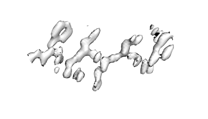
- EMDB-21203: 1.4A damaged structure of GSNQNNF used to determine initial phase... -
+ Open data

- Basic information
| Entry | Database: EMDB / ID: EMD-21203 | |||||||||
|---|---|---|---|---|---|---|---|---|---|---|
| Title | 1.4A damaged structure of GSNQNNF used to determine initial phases from radiation damage | |||||||||
 Map data Map data | 2mFo-Fc map for the damaged GSNQNNF structure refined from the solved low-dose structure that had initially determined experimental phases | |||||||||
 Sample Sample |
| |||||||||
 Keywords Keywords | MicroED / damage / phasing / RIP / protein fibril | |||||||||
| Biological species | synthetic construct (others) | |||||||||
| Method | electron crystallography / cryo EM | |||||||||
 Authors Authors | Martynowycz MW / Hattne J | |||||||||
 Citation Citation | Journal: Structure / Year: 2020 Title: Experimental Phasing of MicroED Data Using Radiation Damage. Authors: Michael W Martynowycz / Johan Hattne / Tamir Gonen /  Abstract: We previously demonstrated that microcrystal electron diffraction (MicroED) can be used to determine atomic-resolution structures from vanishingly small three-dimensional crystals. Here, we present ...We previously demonstrated that microcrystal electron diffraction (MicroED) can be used to determine atomic-resolution structures from vanishingly small three-dimensional crystals. Here, we present an example of an experimentally phased structure using only MicroED data. The structure of a seven-residue peptide is solved starting from differences to the diffraction intensities induced by structural changes due to radiation damage. The same wedge of reciprocal space was recorded twice by continuous-rotation MicroED from a set of 11 individual crystals. The data from the first pass were merged to make a "low-dose dataset." The data from the second pass were similarly merged to form a "damaged dataset." Differences between these two datasets were used to identify a single heavy-atom site from a Patterson difference map, and initial phases were generated. Finally, the structure was completed by iterative cycles of modeling and refinement. | |||||||||
| History |
|
- Structure visualization
| Movie |
 Movie viewer Movie viewer |
|---|---|
| Structure viewer | EM map: SurfViewMolmilJmol/JSmol |
| Supplemental images |
- Downloads & links
-EMDB archive
| Map data | emd_21203.map.gz | 157.4 KB | EMDB map data format | |
|---|---|---|---|---|
| Header (meta data) | emd-21203-v30.xmlemd-21203.xml | 13.3 KB 13.3 KB | Display Display | EMDB header |
| Images |  emd_21203.png emd_21203.png | 82.8 KB | ||
| Filedesc metadata | emd-21203.cif.gz | 5 KB | ||
| Filedesc structureFactors | emd_21203_sf.cif.gz | 59.4 KB | ||
| Archive directory |  http://ftp.pdbj.org/pub/emdb/structures/EMD-21203ftp://ftp.pdbj.org/pub/emdb/structures/EMD-21203 http://ftp.pdbj.org/pub/emdb/structures/EMD-21203ftp://ftp.pdbj.org/pub/emdb/structures/EMD-21203 | HTTPS FTP |
-Related structure data

-Links
| EMDB pages | EMDB (EBI/PDBe) / EMDataResource |
|---|---|
| Related items in Molecule of the Month |

-Map
| File | Download / File: emd_21203.map.gz / Format: CCP4 / Size: 3.3 MB / Type: IMAGE STORED AS FLOATING POINT NUMBER (4 BYTES) | ||||||||||||||||||||||||||||||||||||||||||||||||||||||||||||||||||||
|---|---|---|---|---|---|---|---|---|---|---|---|---|---|---|---|---|---|---|---|---|---|---|---|---|---|---|---|---|---|---|---|---|---|---|---|---|---|---|---|---|---|---|---|---|---|---|---|---|---|---|---|---|---|---|---|---|---|---|---|---|---|---|---|---|---|---|---|---|---|
| Annotation | 2mFo-Fc map for the damaged GSNQNNF structure refined from the solved low-dose structure that had initially determined experimental phases | ||||||||||||||||||||||||||||||||||||||||||||||||||||||||||||||||||||
| Projections & slices | Image control
Images are generated by Spider. generated in cubic-lattice coordinate | ||||||||||||||||||||||||||||||||||||||||||||||||||||||||||||||||||||
| Voxel size | X: 0.40667 Å / Y: 0.44281 Å / Z: 0.4405 Å | ||||||||||||||||||||||||||||||||||||||||||||||||||||||||||||||||||||
| Density |
| ||||||||||||||||||||||||||||||||||||||||||||||||||||||||||||||||||||
| Symmetry | Space group: 1 | ||||||||||||||||||||||||||||||||||||||||||||||||||||||||||||||||||||
| Details | EMDB XML:
CCP4 map header:
| ||||||||||||||||||||||||||||||||||||||||||||||||||||||||||||||||||||
 Z (Sec.)
 X (Row.)
 Y (Col.)


-Supplemental data
- Sample components
-Entire : Synthetic proto-filament
| Entire | Name: Synthetic proto-filament |
|---|---|
| Components |
|
-Supramolecule #1: Synthetic proto-filament
| Supramolecule | Name: Synthetic proto-filament / type: complex / ID: 1 / Parent: 0 / Macromolecule list: #1 |
|---|---|
| Source (natural) | Organism: synthetic construct (others) |
| Molecular weight | Theoretical: 899.141 Da |
-Macromolecule #1: GSNQNNF
| Macromolecule | Name: GSNQNNF / type: protein_or_peptide / ID: 1 / Number of copies: 1 / Enantiomer: LEVO |
|---|---|
| Source (natural) | Organism: synthetic construct (others) |
| Molecular weight | Theoretical: 779.756 Da |
| Sequence | String: GSNQNNF |
-Macromolecule #2: ZINC ION
| Macromolecule | Name: ZINC ION / type: ligand / ID: 2 / Number of copies: 1 / Formula: ZN |
|---|---|
| Molecular weight | Theoretical: 65.409 Da |
-Macromolecule #3: ACETATE ION
| Macromolecule | Name: ACETATE ION / type: ligand / ID: 3 / Number of copies: 1 / Formula: ACT |
|---|---|
| Molecular weight | Theoretical: 59.044 Da |
| Chemical component information |  ChemComp-ACT: |
-Macromolecule #4: water
| Macromolecule | Name: water / type: ligand / ID: 4 / Number of copies: 1 / Formula: HOH |
|---|---|
| Molecular weight | Theoretical: 18.015 Da |
| Chemical component information |  ChemComp-HOH: |
-Experimental details
-Structure determination
| Method | cryo EM |
|---|---|
 Processing Processing | electron crystallography |
| Aggregation state | 3D array |
-Sample preparation
| Concentration | 10 mg/mL | |||||||||
|---|---|---|---|---|---|---|---|---|---|---|
| Buffer | pH: 6 Component:
| |||||||||
| Grid | Model: Quantifoil R2/4 / Material: COPPER / Mesh: 300 / Support film - Material: CARBON / Support film - topology: HOLEY ARRAY / Pretreatment - Type: GLOW DISCHARGE | |||||||||
| Vitrification | Cryogen name: ETHANE / Chamber humidity: 30 % / Instrument: FEI VITROBOT MARK IV | |||||||||
| Details | Hanging drop. |
- Electron microscopy
| Microscope | FEI TECNAI F20 |
|---|---|
| Image recording | Film or detector model: TVIPS TEMCAM-F416 (4k x 4k) / Digitization - Dimensions - Width: 2048 pixel / Digitization - Dimensions - Height: 2048 pixel / Number grids imaged: 1 / Number real images: 736 / Number diffraction images: 736 / Average exposure time: 2.1 sec. / Average electron dose: 0.00588 e/Å2 / Details: Images collected as a movies. |
| Electron beam | Acceleration voltage: 200 kV / Electron source:  FIELD EMISSION GUN FIELD EMISSION GUN |
| Electron optics | C2 aperture diameter: 100.0 µm / Illumination mode: FLOOD BEAM / Imaging mode: DIFFRACTION / Camera length: 730 mm |
| Sample stage | Specimen holder model: GATAN 626 SINGLE TILT LIQUID NITROGEN CRYO TRANSFER HOLDER Cooling holder cryogen: NITROGEN |
| Experimental equipment |  Model: Tecnai F20 / Image courtesy: FEI Company |
-Image processing
| Details | Rolling shutter and binned by 2. |
|---|---|
| Final reconstruction | Resolution method: DIFFRACTION PATTERN/LAYERLINES |
| Crystallography statistics | Number intensities measured: 6314 / Number structure factors: 722 / Fourier space coverage: 78.1 / R sym: 0.21 / R merge: 0.199 / Overall phase error: 26 / Overall phase residual: 26 / Phase error rejection criteria: 0 / High resolution: 1.4 Å Details: Model from the low-dose set refined against the damage dataset without any changes. Shell - Shell ID: 1 / Shell - High resolution: 1.4 Å / Shell - Low resolution: 13.97 Å / Shell - Number structure factors: 722 / Shell - Phase residual: 26 / Shell - Fourier space coverage: 78.1 / Shell - Multiplicity: 8.7 |
-Atomic model buiding 1
| Initial model | PDB ID: Chain - Chain ID: A / Chain - Residue range: 1-7 / Chain - Source name: PDB / Chain - Initial model type: experimental model |
|---|---|
| Refinement | Space: RECIPROCAL / Protocol: OTHER / Overall B value: 5 |
| Output model |  PDB-6vhc: |