[English] 日本語
Yorodumi
- EMDB-20333: CryoEM Plasmodium falciparum M18 aspartyl aminopeptidase -

+
Open data


ID or keywords:

Loading...

-
Basic information

Entry
Database: EMDB / ID: EMD-20333
TitleCryoEM Plasmodium falciparum M18 aspartyl aminopeptidase
Map dataPlasmodium falciparum M18
Sample
  • Complex: Plasmodium falciparum M18 aspartyl aminopeptidase
    • Protein or peptide: M18 aspartyl aminopeptidase
  • Ligand: ZINC ION
Function / homologyPeptidase M18 / Peptidase M18, domain 2 / Aminopeptidase I zinc metalloprotease (M18) / aminopeptidase activity / metallopeptidase activity / zinc ion binding / M18 aspartyl aminopeptidase
Function and homology information
Biological speciesPlasmodium falciparum (malaria parasite P. falciparum) / Plasmodium falciparum (isolate NF54) (eukaryote)
Methodsingle particle reconstruction / cryo EM / Resolution: 3.2 Å
AuthorsHo C / Zhou ZH
Funding support United States, 10 items
OrganizationGrant numberCountry
National Institutes of Health/Office of the DirectorS10OD018111 United States
National Institutes of Health/National Center for Research Resources (NIH/NCRR)R01GM071940 United States
National Institutes of Health/National Center for Research Resources (NIH/NCRR)DE025567 United States
National Institutes of Health/National Center for Research Resources (NIH/NCRR)AI094386 United States
National Institutes of Health/National Center for Research Resources (NIH/NCRR)U24GM116792 United States
National Institutes of Health/National Center for Research Resources (NIH/NCRR)S10RR23057 United States
National Science Foundation (NSF, United States)DMR-1548924 United States
National Science Foundation (NSF, United States)DBI-1338135 United States
National Institutes of Health/National Institute Of Allergy and Infectious Diseases (NIH/NIAID)T32 AI007323 United States
National Institutes of Health/National Heart, Lung, and Blood Institute (NIH/NHLBI)K99/R00 HL133453 United States
Citation
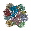
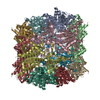
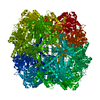
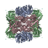
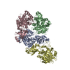
Journal: Nat Methods / Year: 2020
Title: Bottom-up structural proteomics: cryoEM of protein complexes enriched from the cellular milieu.
Authors: Chi-Min Ho / Xiaorun Li / Mason Lai / Thomas C Terwilliger / Josh R Beck / James Wohlschlegel / Daniel E Goldberg / Anthony W P Fitzpatrick / Z Hong Zhou /
Abstract: X-ray crystallography often requires non-native constructs involving mutations or truncations, and is challenged by membrane proteins and large multicomponent complexes. We present here a bottom-up ...X-ray crystallography often requires non-native constructs involving mutations or truncations, and is challenged by membrane proteins and large multicomponent complexes. We present here a bottom-up endogenous structural proteomics approach whereby near-atomic-resolution cryo electron microscopy (cryoEM) maps are reconstructed ab initio from unidentified protein complexes enriched directly from the endogenous cellular milieu, followed by identification and atomic modeling of the proteins. The proteins in each complex are identified using cryoID, a program we developed to identify proteins in ab initio cryoEM maps. As a proof of principle, we applied this approach to the malaria-causing parasite Plasmodium falciparum, an organism that has resisted conventional structural-biology approaches, to obtain atomic models of multiple protein complexes implicated in intraerythrocytic survival of the parasite. Our approach is broadly applicable for determining structures of undiscovered protein complexes enriched directly from endogenous sources.
History
DepositionJun 21, 2019-
Header (metadata) releaseJul 24, 2019-
Map releaseDec 11, 2019-
UpdateJan 15, 2020-
Current statusJan 15, 2020Processing site: RCSB / Status: Released

-
Structure visualization

Movie
  • Surface view with section colored by density value
  • Surface level: 0.108
  • Imaged by UCSF Chimera
  • Download
  • Surface view colored by radius
  • Surface level: 0.108
  • Imaged by UCSF Chimera
  • Download
  • Surface view with fitted model
  • Atomic models: PDB-6pev
  • Surface level: 0.108
  • Imaged by UCSF Chimera
  • Download
Movie viewer
Structure viewerEM map:
SurfViewMolmilJmol/JSmol
Supplemental images

Downloads & links

-
Map

FileDownload / File: emd_20333.map.gz / Format: CCP4 / Size: 125 MB / Type: IMAGE STORED AS FLOATING POINT NUMBER (4 BYTES)
AnnotationPlasmodium falciparum M18
Voxel sizeX=Y=Z: 1.04 Å
Density
Contour LevelBy AUTHOR: 0.108 / Movie #1: 0.108
Minimum - Maximum-0.18047863 - 0.31202805
Average (Standard dev.)0.0005793639 (±0.011046562)
SymmetrySpace group: 1
Details

EMDB XML:

Map geometry
Axis orderXYZ
Origin000
Dimensions320320320
Spacing320320320
CellA=B=C: 332.8 Å
α=β=γ: 90.0 °

CCP4 map header:

modeImage stored as Reals
Å/pix. X/Y/Z1.041.041.04
M x/y/z320320320
origin x/y/z0.0000.0000.000
length x/y/z332.800332.800332.800
α/β/γ90.00090.00090.000
start NX/NY/NZ-200-200-200
NX/NY/NZ401401401
MAP C/R/S123
start NC/NR/NS000
NC/NR/NS320320320
D min/max/mean-0.1800.3120.001

-
Supplemental data

-
Sample components

-
Entire : Plasmodium falciparum M18 aspartyl aminopeptidase

EntireName: Plasmodium falciparum M18 aspartyl aminopeptidase
Components
  • Complex: Plasmodium falciparum M18 aspartyl aminopeptidase
    • Protein or peptide: M18 aspartyl aminopeptidase
  • Ligand: ZINC ION

-
Supramolecule #1: Plasmodium falciparum M18 aspartyl aminopeptidase

SupramoleculeName: Plasmodium falciparum M18 aspartyl aminopeptidase / type: complex / ID: 1 / Parent: 0 / Macromolecule list: #1
Source (natural)Organism: Plasmodium falciparum (malaria parasite P. falciparum)

-
Macromolecule #1: M18 aspartyl aminopeptidase

MacromoleculeName: M18 aspartyl aminopeptidase / type: protein_or_peptide / ID: 1 / Number of copies: 12 / Enantiomer: LEVO
Source (natural)Organism: Plasmodium falciparum (isolate NF54) (eukaryote) / Strain: isolate NF54
Molecular weightTheoretical: 65.72193 KDa
SequenceString: MDKKAREYAQ DALKFIQRSG SNFLACKNLK ERLENNGFIN LSEGETWNLN KNEGYVLCKE NRNICGFFVG KNFNIDTGSI LISIGHIDS CALKISPNNN VIKKKIHQIN VECYGSGLWH TWFDRSLGLS GQVLYKKGNK LVEKLIQINK SVLFLPSLAI H LQNRTRYD ...String:
MDKKAREYAQ DALKFIQRSG SNFLACKNLK ERLENNGFIN LSEGETWNLN KNEGYVLCKE NRNICGFFVG KNFNIDTGSI LISIGHIDS CALKISPNNN VIKKKIHQIN VECYGSGLWH TWFDRSLGLS GQVLYKKGNK LVEKLIQINK SVLFLPSLAI H LQNRTRYD FSVKINYENH IKPIISTTLF NQLNKCKRNN VHHDTILTTD TKFSHKENSQ NKRDDQMCHS FNDKDVSNHN LD KNTIEHL TNQQNEEKNK HTKDNPNSKD IVEHINTDNS YPLLYLLSKE LNCKEEDILD FELCLMDTQE PCFTGVYEEF IEG ARFDNL LGSFCVFEGF IELVNSIKNH TSNENTNHTN NITNDINDNI HNNLYISIGY DHEEIGSLSE VGARSYCTKN FIDR IISSV FKKEIHEKNL SVQEIYGNLV NRSFILNVDM AHCSHPNYPE TVQDNHQLFF HEGIAIKYNT NKNYVTSPLH ASLIK RTFE LYYNKYKQQI KYQNFMVKND TPCGSTVGSM VAANLSMPGI DIGIPQLAMH SIREIAAVHD VFFLIKGVFA FYTYYN QVL STCVHDK

-
Macromolecule #2: ZINC ION

MacromoleculeName: ZINC ION / type: ligand / ID: 2 / Number of copies: 24 / Formula: ZN
Molecular weightTheoretical: 65.409 Da

-
Experimental details

-
Structure determination

Methodcryo EM
Processingsingle particle reconstruction
Aggregation stateparticle

-
Sample preparation

BufferpH: 7.4
VitrificationCryogen name: ETHANE

-
Electron microscopy

MicroscopeFEI TITAN KRIOS
Image recordingFilm or detector model: GATAN K2 SUMMIT (4k x 4k) / Detector mode: COUNTING / Average electron dose: 60.0 e/Å2
Electron beamAcceleration voltage: 300 kV / Electron source: FIELD EMISSION GUN
Electron opticsC2 aperture diameter: 50.0 µm / Calibrated defocus max: 4.0 µm / Calibrated defocus min: 1.5 µm / Illumination mode: FLOOD BEAM / Imaging mode: BRIGHT FIELD / Cs: 2.7 mm
Experimental equipment
Model: Titan Krios / Image courtesy: FEI Company

-
Image processing

CTF correctionSoftware - Name: cryoSPARC (ver. 1)
Final reconstructionNumber classes used: 1 / Applied symmetry - Point group: T (tetrahedral) / Resolution.type: BY AUTHOR / Resolution: 3.2 Å / Resolution method: FSC 0.143 CUT-OFF / Software - Name: cryoSPARC (ver. 1) / Number images used: 5860
Initial angle assignmentType: OTHER / Software - Name: cryoSPARC (ver. 1)
Final angle assignmentType: OTHER / Software - Name: cryoSPARC (ver. 1)

+
About Yorodumi

-
News

-
Feb 9, 2022. New format data for meta-information of EMDB entries

New format data for meta-information of EMDB entries

  • Version 3 of the EMDB header file is now the official format.
  • The previous official version 1.9 will be removed from the archive.

Related info.:EMDB header

External links:wwPDB to switch to version 3 of the EMDB data model

-
Aug 12, 2020. Covid-19 info

Covid-19 info

URL: https://pdbjlc1.pdbj.org/emnavi/covid19.php

New page: Covid-19 featured information page in EM Navigator.

Related info.:Covid-19 info / Mar 5, 2020. Novel coronavirus structure data

+
Mar 5, 2020. Novel coronavirus structure data

Novel coronavirus structure data

Related info.:Yorodumi Speices / Aug 12, 2020. Covid-19 info

External links:COVID-19 featured content - PDBj / Molecule of the Month (242):Coronavirus Proteases

+
Jan 31, 2019. EMDB accession codes are about to change! (news from PDBe EMDB page)

EMDB accession codes are about to change! (news from PDBe EMDB page)

  • The allocation of 4 digits for EMDB accession codes will soon come to an end. Whilst these codes will remain in use, new EMDB accession codes will include an additional digit and will expand incrementally as the available range of codes is exhausted. The current 4-digit format prefixed with “EMD-” (i.e. EMD-XXXX) will advance to a 5-digit format (i.e. EMD-XXXXX), and so on. It is currently estimated that the 4-digit codes will be depleted around Spring 2019, at which point the 5-digit format will come into force.
  • The EM Navigator/Yorodumi systems omit the EMD- prefix.

Related info.:Q: What is EMD? / ID/Accession-code notation in Yorodumi/EM Navigator

External links:EMDB Accession Codes are Changing Soon! / Contact to PDBj

+
Jul 12, 2017. Major update of PDB

Major update of PDB

  • wwPDB released updated PDB data conforming to the new PDBx/mmCIF dictionary.
  • This is a major update changing the version number from 4 to 5, and with Remediation, in which all the entries are updated.
  • In this update, many items about electron microscopy experimental information are reorganized (e.g. em_software).
  • Now, EM Navigator and Yorodumi are based on the updated data.

External links:wwPDB Remediation / Enriched Model Files Conforming to OneDep Data Standards Now Available in the PDB FTP Archive

-
Yorodumi

Thousand views of thousand structures

  • Yorodumi is a browser for structure data from EMDB, PDB, SASBDB, etc.
  • This page is also the successor to EM Navigator detail page, and also detail information page/front-end page for Omokage search.
  • The word "yorodu" (or yorozu) is an old Japanese word meaning "ten thousand". "mi" (miru) is to see.

Related info.:EMDB / PDB / SASBDB / Comparison of 3 databanks / Yorodumi Search / Aug 31, 2016. New EM Navigator & Yorodumi / Yorodumi Papers / Jmol/JSmol / Function and homology information / Changes in new EM Navigator and Yorodumi

Read more