 Movie
Movie Controller

[English] 日本語
 Yorodumi
- EMDB-2004: Asymmetric reconstruction of 13-protofilament GTPgammaS microtubu... -
+ Open data

- Basic information
| Entry | Database: EMDB / ID: EMD-2004 | |||||||||
|---|---|---|---|---|---|---|---|---|---|---|
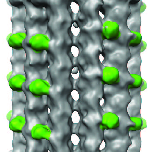
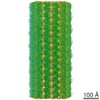
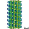
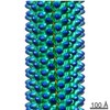
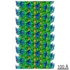
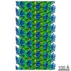
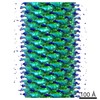
| Title | Asymmetric reconstruction of 13-protofilament GTPgammaS microtubules decorated with Mal3 CH domain | |||||||||
 Map data Map data | Asymmetric reconstruction of 13-protofilament GTPgammaS microtubules decorated with Mal3 CH domain | |||||||||
 Sample Sample |
| |||||||||
 Keywords Keywords | cytoskeleton / GTPase / end binding / calponin homology | |||||||||
| Biological species |  | |||||||||
| Method | single particle reconstruction / cryo EM / Resolution: 15.0 Å | |||||||||
 Authors Authors | Maurer SP / Fourniol FJ / Bohner G / Moores CA / Surrey T | |||||||||
 Citation Citation | Journal: Cell / Year: 2012 Title: EBs recognize a nucleotide-dependent structural cap at growing microtubule ends. Authors: Sebastian P Maurer / Franck J Fourniol / Gergő Bohner / Carolyn A Moores / Thomas Surrey /  Abstract: Growing microtubule ends serve as transient binding platforms for essential proteins that regulate microtubule dynamics and their interactions with cellular substructures. End-binding proteins (EBs) ...Growing microtubule ends serve as transient binding platforms for essential proteins that regulate microtubule dynamics and their interactions with cellular substructures. End-binding proteins (EBs) autonomously recognize an extended region at growing microtubule ends with unknown structural characteristics and then recruit other factors to the dynamic end structure. Using cryo-electron microscopy, subnanometer single-particle reconstruction, and fluorescence imaging, we present a pseudoatomic model of how the calponin homology (CH) domain of the fission yeast EB Mal3 binds to the end regions of growing microtubules. The Mal3 CH domain bridges protofilaments except at the microtubule seam. By binding close to the exchangeable GTP-binding site, the CH domain is ideally positioned to sense the microtubule's nucleotide state. The same microtubule-end region is also a stabilizing structural cap protecting the microtubule from depolymerization. This insight supports a common structural link between two important biological phenomena, microtubule dynamic instability and end tracking. | |||||||||
| History |
|
- Structure visualization
| Movie |
 Movie viewer Movie viewer |
|---|---|
| Structure viewer | EM map: SurfViewMolmilJmol/JSmol |
| Supplemental images |
- Downloads & links
-EMDB archive
| Map data | emd_2004.map.gz | 56.5 MB | EMDB map data format | |
|---|---|---|---|---|
| Header (meta data) | emd-2004-v30.xmlemd-2004.xml | 10.3 KB 10.3 KB | Display Display | EMDB header |
| Images |  2004.jpg 2004.jpg | 846 KB | ||
| Archive directory |  http://ftp.pdbj.org/pub/emdb/structures/EMD-2004ftp://ftp.pdbj.org/pub/emdb/structures/EMD-2004 http://ftp.pdbj.org/pub/emdb/structures/EMD-2004ftp://ftp.pdbj.org/pub/emdb/structures/EMD-2004 | HTTPS FTP |
-Related structure data

-Links
| EMDB pages | EMDB (EBI/PDBe) / EMDataResource |
|---|
-Map
| File | Download / File: emd_2004.map.gz / Format: CCP4 / Size: 73.3 MB / Type: IMAGE STORED AS FLOATING POINT NUMBER (4 BYTES) | ||||||||||||||||||||||||||||||||||||||||||||||||||||||||||||||||||||
|---|---|---|---|---|---|---|---|---|---|---|---|---|---|---|---|---|---|---|---|---|---|---|---|---|---|---|---|---|---|---|---|---|---|---|---|---|---|---|---|---|---|---|---|---|---|---|---|---|---|---|---|---|---|---|---|---|---|---|---|---|---|---|---|---|---|---|---|---|---|
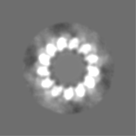
| Annotation | Asymmetric reconstruction of 13-protofilament GTPgammaS microtubules decorated with Mal3 CH domain | ||||||||||||||||||||||||||||||||||||||||||||||||||||||||||||||||||||
| Projections & slices | Image control
Images are generated by Spider. | ||||||||||||||||||||||||||||||||||||||||||||||||||||||||||||||||||||
| Voxel size | X=Y=Z: 2.2 Å | ||||||||||||||||||||||||||||||||||||||||||||||||||||||||||||||||||||
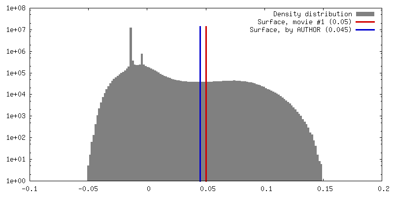
| Density |
| ||||||||||||||||||||||||||||||||||||||||||||||||||||||||||||||||||||
| Symmetry | Space group: 1 | ||||||||||||||||||||||||||||||||||||||||||||||||||||||||||||||||||||
| Details | EMDB XML:
CCP4 map header:
| ||||||||||||||||||||||||||||||||||||||||||||||||||||||||||||||||||||
 Z (Sec.)
 Y (Row.)
 X (Col.)


-Supplemental data
- Sample components
-Entire : 13-protofilament GTPgammaS microtubule decorated with monomeric Mal3
| Entire | Name: 13-protofilament GTPgammaS microtubule decorated with monomeric Mal3 |
|---|---|
| Components |
|
-Supramolecule #1000: 13-protofilament GTPgammaS microtubule decorated with monomeric Mal3
| Supramolecule | Name: 13-protofilament GTPgammaS microtubule decorated with monomeric Mal3 type: sample / ID: 1000 Details: tubulin was mixed with GMPCPP microtubule seeds, GTPgammaS and monomeric Mal3, and incubated 3-10min at 37degC Oligomeric state: 13-protofilament microtubule / Number unique components: 3 |
|---|
-Macromolecule #1: Alpha-beta tubulin dimer
| Macromolecule | Name: Alpha-beta tubulin dimer / type: protein_or_peptide / ID: 1 / Name.synonym: Alpha-beta tubulin dimer / Oligomeric state: Dimer / Recombinant expression: No |
|---|---|
| Source (natural) | Organism: |
| Molecular weight | Experimental: 100 KDa / Theoretical: 100 KDa |
-Macromolecule #3: Mal3
| Macromolecule | Name: Mal3 / type: protein_or_peptide / ID: 3 / Name.synonym: Mal3 / Oligomeric state: monomer / Recombinant expression: Yes |
|---|---|
| Source (natural) | Organism: Saccharomyces pombe / synonym: fission yeast / Location in cell: cytoplasmic |
| Molecular weight | Experimental: 16 KDa / Theoretical: 16 KDa |
| Recombinant expression | Organism:  |
-Macromolecule #2: guanosine 5'-O-(gamma-thio)triphosphate
| Macromolecule | Name: guanosine 5'-O-(gamma-thio)triphosphate / type: ligand / ID: 2 / Name.synonym: GTPgammaS / Recombinant expression: No |
|---|---|
| Source (natural) | Organism: synthetic construct (others) |
-Experimental details
-Structure determination
| Method | cryo EM |
|---|---|
 Processing Processing | single particle reconstruction |
| Aggregation state | particle |
-Sample preparation
| Buffer | pH: 6.8 / Details: 40mM Pipes, 1mM MgCl2, 1mM EGTA |
|---|---|
| Grid | Details: 300 mesh lacey carbon grid |
| Vitrification | Cryogen name: ETHANE / Chamber humidity: 90 % / Instrument: OTHER / Details: Vitrification instrument: Vitrobot (FEI) / Method: Chamber at 37 degrees C, blot 2s |
- Electron microscopy
| Microscope | FEI TECNAI F20 |
|---|---|
| Temperature | Min: 88 K / Max: 98 K / Average: 93 K |
| Alignment procedure | Legacy - Astigmatism: Objective lens astigmatism was corrected at 150,000 times magnification |
| Image recording | Category: CCD / Film or detector model: GATAN ULTRASCAN 4000 (4k x 4k) / Number real images: 162 / Average electron dose: 17 e/Å2 / Details: sampling size 2.2 A per pixel |
| Electron beam | Acceleration voltage: 200 kV / Electron source:  FIELD EMISSION GUN FIELD EMISSION GUN |
| Electron optics | Illumination mode: FLOOD BEAM / Imaging mode: BRIGHT FIELD / Cs: 2.0 mm / Nominal defocus max: 3.6 µm / Nominal defocus min: 0.7 µm / Nominal magnification: 68000 |
| Sample stage | Specimen holder: Eucentric / Specimen holder model: OTHER |
| Experimental equipment |  Model: Tecnai F20 / Image courtesy: FEI Company |
-Image processing
| CTF correction | Details: FREALIGN |
|---|---|
| Final reconstruction | Applied symmetry - Point group: C1 (asymmetric) / Algorithm: OTHER / Resolution.type: BY AUTHOR / Resolution: 15.0 Å / Resolution method: FSC 0.5 CUT-OFF / Software - Name: SPIDER, FREALIGN Details: C1 map calculated from approximately 11000 one-dimer-long microtubule segments |
