 Movie
Movie Controller

[English] 日本語
 Yorodumi
- PDB-1ytz: Crystal structure of skeletal muscle troponin in the Ca2+-activat... -
+ Open data

- Basic information
| Entry | Database: PDB / ID: 1ytz | ||||||
|---|---|---|---|---|---|---|---|
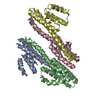
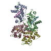
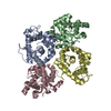
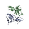
| Title | Crystal structure of skeletal muscle troponin in the Ca2+-activated state | ||||||
 Components Components |
| ||||||
 Keywords Keywords | CONTRACTILE PROTEIN / troponin / muscle / thin filament / actin binding / calcium | ||||||
| Function / homology |  Function and homology information Function and homology informationtroponin C binding / Striated Muscle Contraction / troponin complex / regulation of muscle contraction / tropomyosin binding / troponin I binding / skeletal muscle contraction / cardiac muscle contraction / actin binding / calcium ion binding Similarity search - Function | ||||||
| Biological species |  | ||||||
| Method |  X-RAY DIFFRACTION / SYNCHROTRON / MOLECULAR REPLACEMENT / Resolution: 3 Å X-RAY DIFFRACTION / SYNCHROTRON / MOLECULAR REPLACEMENT / Resolution: 3 Å | ||||||
 Authors Authors | Vinogradova, M.V. / Stone, D.B. / Malanina, G.G. / Karatzaferi, C. / Cooke, R. / Mendelson, R.A. / Fletterick, R.J. | ||||||
 Citation Citation | Journal: Proc.Natl.Acad.Sci.Usa / Year: 2005 Title: Ca2+-regulated structural changes in troponin Authors: Vinogradova, M.V. / Stone, D.B. / Malanina, G.G. / Karatzaferi, C. / Cooke, R. / Mendelson, R.A. / Fletterick, R.J. | ||||||
| History |
|
- Structure visualization
| Structure viewer | Molecule: MolmilJmol/JSmol |
|---|
- Downloads & links
-Download
| PDBx/mmCIF format | 1ytz.cif.gz | 101.5 KB | Display | PDBx/mmCIF format |
|---|---|---|---|---|
| PDB format | pdb1ytz.ent.gz | 73.3 KB | Display | PDB format |
| PDBx/mmJSON format | 1ytz.json.gz | Tree view | PDBx/mmJSON format | |
| Others |  Other downloads Other downloads |
-Validation report
| Summary document | 1ytz_validation.pdf.gz | 937.6 KB | Display | wwPDB validaton report |
|---|---|---|---|---|
| Full document | 1ytz_full_validation.pdf.gz | 962.5 KB | Display | |
| Data in XML | 1ytz_validation.xml.gz | 20.1 KB | Display | |
| Data in CIF | 1ytz_validation.cif.gz | 26.1 KB | Display | |
| Arichive directory | https://data.pdbj.org/pub/pdb/validation_reports/yt/1ytzftp://data.pdbj.org/pub/pdb/validation_reports/yt/1ytz | HTTPS FTP |
-Related structure data


-Links
 PDBj
PDBj

- Assembly
| Deposited unit | 
| ||||||||
|---|---|---|---|---|---|---|---|---|---|
| 1 |
| ||||||||
| 2 | 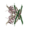
| ||||||||
| Unit cell |
|
-Components
-Protein , 3 types, 3 molecules TIC
| #1: Protein | Mass: 12694.692 Da / Num. of mol.: 1 Source method: isolated from a genetically manipulated source Source: (gene. exp.)  |
|---|---|
| #2: Protein | Mass: 21116.246 Da / Num. of mol.: 1 Source method: isolated from a genetically manipulated source Source: (gene. exp.) |
| #3: Protein | Mass: 18261.188 Da / Num. of mol.: 1 Source method: isolated from a genetically manipulated source Source: (gene. exp.) |
-Non-polymers , 3 types, 40 molecules 


| #4: Chemical |  Mass: 1527.901 Da / Num. of mol.: 3 / Source method: obtained synthetically / Formula: C74H142O31 Mass: 1527.901 Da / Num. of mol.: 3 / Source method: obtained synthetically / Formula: C74H142O31#5: Chemical | ChemComp-CA /  Mass: 40.078 Da / Num. of mol.: 4 / Source method: obtained synthetically / Formula: Ca Mass: 40.078 Da / Num. of mol.: 4 / Source method: obtained synthetically / Formula: Ca#6: Water | ChemComp-HOH / | Mass: 18.015 Da / Num. of mol.: 33 / Source method: isolated from a natural source / Formula: H2O |
|---|
-Experimental details
-Experiment
| Experiment | Method: X-RAY DIFFRACTION / Number of used crystals: 1 |
|---|
- Sample preparation
| Crystal | Density Matthews: 3.864 Å3/Da / Density % sol: 67 % |
|---|---|
| Crystal grow | Temperature: 298 K / Method: vapor diffusion, sitting drop / pH: 8 Details: 0.4 M NaH2PO4/1.6 M K2HPO4, 200 mM NaCl, 0.1 M immidazole, pH 8.0, VAPOR DIFFUSION, SITTING DROP, temperature 298K |
-Data collection
| Diffraction | Mean temperature: 100 K |
|---|---|
| Diffraction source | Source: SYNCHROTRON / Site: ALS  / Beamline: 8.3.1 / Wavelength: 1.1 Å / Beamline: 8.3.1 / Wavelength: 1.1 Å |
| Detector | Type: ADSC QUANTUM 4 / Detector: CCD / Date: May 22, 2002 |
| Radiation | Monochromator: double crystal / Protocol: SINGLE WAVELENGTH / Monochromatic (M) / Laue (L): M / Scattering type: x-ray |
| Radiation wavelength | Wavelength: 1.1 Å / Relative weight: 1 |
| Reflection | Resolution: 3→25 Å / Num. all: 16801 / Num. obs: 16095 / % possible obs: 95.9 % / Observed criterion σ(F): 0 / Observed criterion σ(I): 0 / Rmerge(I) obs: 0.055 |
| Reflection shell | Resolution: 3→3.11 Å / % possible all: 90.8 |
- Processing
| Software |
| |||||||||||||||||||||||||||||||||||||||||||||||||
|---|---|---|---|---|---|---|---|---|---|---|---|---|---|---|---|---|---|---|---|---|---|---|---|---|---|---|---|---|---|---|---|---|---|---|---|---|---|---|---|---|---|---|---|---|---|---|---|---|---|---|
| Refinement | Method to determine structure: MOLECULAR REPLACEMENT / Resolution: 3→25 Å / σ(F): 0 / Stereochemistry target values: Engh & Huber
| |||||||||||||||||||||||||||||||||||||||||||||||||
| Refinement step | Cycle: LAST / Resolution: 3→25 Å
| |||||||||||||||||||||||||||||||||||||||||||||||||
| Refine LS restraints |
| |||||||||||||||||||||||||||||||||||||||||||||||||
| LS refinement shell |
|
