 Movie
Movie Controller

[English] 日本語
 Yorodumi
- PDB-1xsp: Crystal Structure of human DNA polymerase lambda in complex with ... -
+ Open data

- Basic information
| Entry | Database: PDB / ID: 1xsp | ||||||
|---|---|---|---|---|---|---|---|
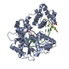
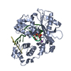
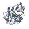
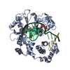
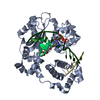
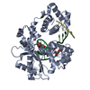
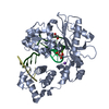
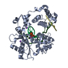
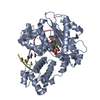
| Title | Crystal Structure of human DNA polymerase lambda in complex with nicked DNA and pyrophosphate | ||||||
 Components Components |
| ||||||
 Keywords Keywords | TRANSFERASE/DNA / DNA POLYMERASE LAMBDA / Protein-DNA complex / Helix-hairpin-helix / TRANSFERASE-DNA COMPLEX | ||||||
| Function / homology |  Function and homology information Function and homology informationDNA biosynthetic process / Lyases; Carbon-oxygen lyases; Other carbon-oxygen lyases / 5'-deoxyribose-5-phosphate lyase activity / somatic hypermutation of immunoglobulin genes / base-excision repair, gap-filling / nucleotide-excision repair / Nonhomologous End-Joining (NHEJ) / double-strand break repair via homologous recombination / double-strand break repair via nonhomologous end joining / site of double-strand break ...DNA biosynthetic process / Lyases; Carbon-oxygen lyases; Other carbon-oxygen lyases / 5'-deoxyribose-5-phosphate lyase activity / somatic hypermutation of immunoglobulin genes / base-excision repair, gap-filling / nucleotide-excision repair / Nonhomologous End-Joining (NHEJ) / double-strand break repair via homologous recombination / double-strand break repair via nonhomologous end joining / site of double-strand break / DNA-directed DNA polymerase / DNA-directed DNA polymerase activity / DNA replication / DNA binding / nucleoplasm / metal ion binding / nucleus Similarity search - Function | ||||||
| Biological species |  Homo sapiens (human) Homo sapiens (human) | ||||||
| Method |  X-RAY DIFFRACTION / MOLECULAR REPLACEMENT / Resolution: 2.2 Å X-RAY DIFFRACTION / MOLECULAR REPLACEMENT / Resolution: 2.2 Å | ||||||
 Authors Authors | Garcia-Diaz, M. / Bebenek, K. / Krahn, J.M. / Kunkel, T.A. / Pedersen, L.C. | ||||||
 Citation Citation | Journal: Nat.Struct.Mol.Biol. / Year: 2005 Title: A closed conformation for the Pol lambda catalytic cycle. Authors: Garcia-Diaz, M. / Bebenek, K. / Krahn, J.M. / Kunkel, T.A. / Pedersen, L.C. #1: Journal: Mol.Cell / Year: 2004Title: A Structural Solution for the DNA Polymerase-lambda Dependent Repair of DNA Gaps with Minimal Homology Authors: Garcia-Diaz, M. / Bebenek, K. / Krahn, J.M. / Blanco, L. / Kunkel, T.A. / Pedersen, L.C. #2: Journal: J.Biol.Chem. / Year: 2002Title: DNA polymerase lambda, a novel DNA repair enzyme in human cells Authors: Garcia-Diaz, M. / Bebenek, K. / Sabariegos, R. / Dominguez, O. / Rodriguez, J. / Kirchhoff, T. / Garcia-Palomero, E. / Picher, A.J. / Juarez, R. / Ruiz, J.F. / Kunkel, T.A. / Blanco, L. #3: Journal: J.Biol.Chem. / Year: 2003Title: The frameshift Infidelity of human DNA polymerase lambda. Implications for function. Authors: Bebenek, K. / Garcia-Diaz, K. / Blanco, L. / Kunkel, T.A. #4: Journal: J.Biol.Chem. / Year: 2001Title: Identification of an intrinsic 5'-deoxyribose-5-phosphate lyase activity in human DNA polymerase lambda: a possible role in Base Excision Repair Authors: Garcia-Diaz, M. / Bebenek, K. / Kunkel, T.A. / Blanco, L. | ||||||
| History |
|
- Structure visualization
| Structure viewer | Molecule: MolmilJmol/JSmol |
|---|
- Downloads & links
-Download
| PDBx/mmCIF format | 1xsp.cif.gz | 103.4 KB | Display | PDBx/mmCIF format |
|---|---|---|---|---|
| PDB format | pdb1xsp.ent.gz | 73.8 KB | Display | PDB format |
| PDBx/mmJSON format | 1xsp.json.gz | Tree view | PDBx/mmJSON format | |
| Others |  Other downloads Other downloads |
-Validation report
| Arichive directory | https://data.pdbj.org/pub/pdb/validation_reports/xs/1xspftp://data.pdbj.org/pub/pdb/validation_reports/xs/1xsp | HTTPS FTP |
|---|
-Related structure data


-Links
 PDBj
PDBj


- Assembly
| Deposited unit | 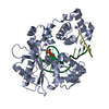
| ||||||||
|---|---|---|---|---|---|---|---|---|---|
| 1 |
| ||||||||
| Unit cell |
| ||||||||
| Details | The protein is a monomer thus the asymmetric unit represents the biological unit |
-Components
-DNA chain , 3 types, 3 molecules TPD
| #1: DNA chain | Mass: 3350.185 Da / Num. of mol.: 1 / Source method: obtained synthetically / Details: Template DNA |
|---|---|
| #2: DNA chain | Mass: 2122.424 Da / Num. of mol.: 1 / Source method: obtained synthetically / Details: Primer DNA |
| #3: DNA chain | Mass: 1191.818 Da / Num. of mol.: 1 / Source method: obtained synthetically / Details: Downstream Primer DNA |
-Protein , 1 types, 1 molecules A
| #4: Protein | Mass: 37447.688 Da / Num. of mol.: 1 / Fragment: 39 kDa catalytic C-terminal domain / Mutation: C543A Source method: isolated from a genetically manipulated source Source: (gene. exp.) Homo sapiens (human) / Gene: POLL / Plasmid: pET22-b / Production host:  |
|---|
-Non-polymers , 3 types, 339 molecules 

| #5: Chemical | ChemComp-NA /  Mass: 22.990 Da / Num. of mol.: 4 / Source method: obtained synthetically / Formula: Na Mass: 22.990 Da / Num. of mol.: 4 / Source method: obtained synthetically / Formula: Na#6: Chemical | ChemComp-PPV / |  Mass: 177.975 Da / Num. of mol.: 1 / Source method: obtained synthetically / Formula: H4O7P2 Mass: 177.975 Da / Num. of mol.: 1 / Source method: obtained synthetically / Formula: H4O7P2#7: Water | ChemComp-HOH / | Mass: 18.015 Da / Num. of mol.: 334 / Source method: isolated from a natural source / Formula: H2O |
|---|
-Experimental details
-Experiment
| Experiment | Method: X-RAY DIFFRACTION / Number of used crystals: 1 |
|---|
- Sample preparation
| Crystal | Density Matthews: 2.7 Å3/Da / Density % sol: 54 % | ||||||||||||||||||||
|---|---|---|---|---|---|---|---|---|---|---|---|---|---|---|---|---|---|---|---|---|---|
| Crystal grow | Temperature: 277 K / Method: vapor diffusion, hanging drop / pH: 5.5 Details: 2-propanol, sodium citrate, sodium chloride, cacodylate, pH 5.5, VAPOR DIFFUSION, HANGING DROP, temperature 277K | ||||||||||||||||||||
| Components of the solutions |
|
-Data collection
| Diffraction | Mean temperature: 100 K |
|---|---|
| Diffraction source | Source: ROTATING ANODE / Type: RIGAKU RUH3R / Wavelength: 1.5418 Å |
| Detector | Type: RIGAKU RAXIS IV / Detector: IMAGE PLATE / Date: Jun 12, 2004 |
| Radiation | Protocol: SINGLE WAVELENGTH / Monochromatic (M) / Laue (L): M / Scattering type: x-ray |
| Radiation wavelength | Wavelength: 1.5418 Å / Relative weight: 1 |
| Reflection | Resolution: 2.2→50 Å / Num. obs: 25700 / % possible obs: 98.6 % / Observed criterion σ(F): 0 / Observed criterion σ(I): -3 / Redundancy: 6.1 % / Biso Wilson estimate: 29.9 Å2 / Rsym value: 0.114 / Net I/σ(I): 16.23 |
| Reflection shell | Resolution: 2.2→2.28 Å / Redundancy: 3.1 % / % possible all: 91.8 |
- Processing
| Software |
| ||||||||||||||||||||||||||||||||||||||||||||||||||||||||||||||||||||||||||||||||
|---|---|---|---|---|---|---|---|---|---|---|---|---|---|---|---|---|---|---|---|---|---|---|---|---|---|---|---|---|---|---|---|---|---|---|---|---|---|---|---|---|---|---|---|---|---|---|---|---|---|---|---|---|---|---|---|---|---|---|---|---|---|---|---|---|---|---|---|---|---|---|---|---|---|---|---|---|---|---|---|---|---|
| Refinement | Method to determine structure: MOLECULAR REPLACEMENT / Resolution: 2.2→41.72 Å / Rfactor Rfree error: 0.007 / Data cutoff high absF: 141990.86 / Data cutoff low absF: 0 / Isotropic thermal model: RESTRAINED / Cross valid method: THROUGHOUT / σ(F): 0 / Stereochemistry target values: Engh & Huber
| ||||||||||||||||||||||||||||||||||||||||||||||||||||||||||||||||||||||||||||||||
| Solvent computation | Solvent model: FLAT MODEL / Bsol: 43.7143 Å2 / ksol: 0.308968 e/Å3 | ||||||||||||||||||||||||||||||||||||||||||||||||||||||||||||||||||||||||||||||||
| Displacement parameters | Biso mean: 41.9 Å2
| ||||||||||||||||||||||||||||||||||||||||||||||||||||||||||||||||||||||||||||||||
| Refine analyze |
| ||||||||||||||||||||||||||||||||||||||||||||||||||||||||||||||||||||||||||||||||
| Refinement step | Cycle: LAST / Resolution: 2.2→41.72 Å
| ||||||||||||||||||||||||||||||||||||||||||||||||||||||||||||||||||||||||||||||||
| Refine LS restraints |
| ||||||||||||||||||||||||||||||||||||||||||||||||||||||||||||||||||||||||||||||||
| LS refinement shell | Resolution: 2.2→2.34 Å / Rfactor Rfree error: 0.028 / Total num. of bins used: 6
| ||||||||||||||||||||||||||||||||||||||||||||||||||||||||||||||||||||||||||||||||
| Xplor file |
|
