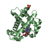
 Movie
Movie Controller

[English] 日本語
 Yorodumi
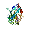
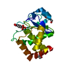
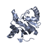
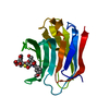
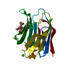
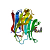
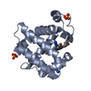
- PDB-1umt: Stromelysin-1 catalytic domain with hydrophobic inhibitor bound, ... -
+ Open data

- Basic information
| Entry | Database: PDB / ID: 1umt | ||||||
|---|---|---|---|---|---|---|---|
| Title | Stromelysin-1 catalytic domain with hydrophobic inhibitor bound, ph 7.0, 32oc, 20 mm cacl2, 15% acetonitrile; nmr average of 20 structures minimized with restraints | ||||||
 Components Components | STROMELYSIN-1 | ||||||
 Keywords Keywords | HYDROLASE/HYDROLASE INHIBITOR / ZINC HYDROLASE / METZINCIN / MATRIX METALLOPROTEINASE / HYDROLASE-HYDROLASE INHIBITOR COMPLEX | ||||||
| Function / homology |  Function and homology information Function and homology informationstromelysin 1 / cellular response to UV-A / regulation of neuroinflammatory response / Assembly of collagen fibrils and other multimeric structures / Activation of Matrix Metalloproteinases / response to amyloid-beta / Collagen degradation / collagen catabolic process / negative regulation of reactive oxygen species metabolic process / extracellular matrix disassembly ...stromelysin 1 / cellular response to UV-A / regulation of neuroinflammatory response / Assembly of collagen fibrils and other multimeric structures / Activation of Matrix Metalloproteinases / response to amyloid-beta / Collagen degradation / collagen catabolic process / negative regulation of reactive oxygen species metabolic process / extracellular matrix disassembly / Degradation of the extracellular matrix / extracellular matrix organization / cellular response to nitric oxide / EGFR Transactivation by Gastrin / regulation of cell migration / negative regulation of phosphatidylinositol 3-kinase/protein kinase B signal transduction / protein catabolic process / cellular response to reactive oxygen species / cellular response to amino acid stimulus / positive regulation of protein-containing complex assembly / metalloendopeptidase activity / metallopeptidase activity / peptidase activity / extracellular matrix / cellular response to lipopolysaccharide / Interleukin-4 and Interleukin-13 signaling / endopeptidase activity / Extra-nuclear estrogen signaling / serine-type endopeptidase activity / innate immune response / mitochondrion / proteolysis / : / extracellular region / zinc ion binding / nucleus / cytosol Similarity search - Function | ||||||
| Biological species |  Homo sapiens (human) Homo sapiens (human) | ||||||
| Method | SOLUTION NMR | ||||||
| Model type details | minimized average | ||||||
 Authors Authors | Van Doren, S.R. / Kurochkin, A.V. / Hu, W. / Zuiderweg, E.R.P. | ||||||
 Citation Citation | Journal: Protein Sci. / Year: 1995 Title: Solution structure of the catalytic domain of human stromelysin complexed with a hydrophobic inhibitor. Authors: Van Doren, S.R. / Kurochkin, A.V. / Hu, W. / Ye, Q.Z. / Johnson, L.L. / Hupe, D.J. / Zuiderweg, E.R. #1: Journal: Biochemistry / Year: 1993Title: Assignments for the Main-Chain Nuclear Magnetic Resonances and Delineation of the Secondary Structure of the Catalytic Domain of Human Stromelysin-1 as Obtained from Triple-Resonance 3D NMR Experiments Authors: Van Doren, S.R. / Kurochkin, A.V. / Ye, Q.-Z. / Johnson, L.L. / Hupe, D.J. / Zuiderweg, E.R.P. | ||||||
| History |
|
- Structure visualization
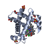
| Structure viewer | Molecule: MolmilJmol/JSmol |
|---|
- Downloads & links
-Download
| PDBx/mmCIF format | 1umt.cif.gz | 69.7 KB | Display | PDBx/mmCIF format |
|---|---|---|---|---|
| PDB format | pdb1umt.ent.gz | 50.2 KB | Display | PDB format |
| PDBx/mmJSON format | 1umt.json.gz | Tree view | PDBx/mmJSON format | |
| Others |  Other downloads Other downloads |
-Validation report
| Arichive directory | https://data.pdbj.org/pub/pdb/validation_reports/um/1umtftp://data.pdbj.org/pub/pdb/validation_reports/um/1umt | HTTPS FTP |
|---|
-Related structure data


-Links
 PDBj
PDBj


- Assembly
| Deposited unit | 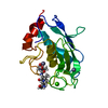
| |||||||||
|---|---|---|---|---|---|---|---|---|---|---|
| 1 |
| |||||||||
| Atom site foot note | 1: CIS PROLINE - PRO A 87 2: THR A 128 - PRO A 129 OMEGA = 137.36 PEPTIDE BOND DEVIATES SIGNIFICANTLY FROM TRANS CONFORMATION | |||||||||
| NMR ensembles |
|
-Components
| #1: Protein | Mass: 19513.646 Da / Num. of mol.: 1 / Fragment: CATALYTIC DOMAIN RESIDUES 83 - 256 Source method: isolated from a genetically manipulated source Source: (gene. exp.) Homo sapiens (human) / Description: INDUCTION BY M13 WITH T7 RNA POLYMERASE / Gene: HUMAN STROMELYSIN-1 CATALYTIC / Plasmid: PGEMEX-DGene (production host): HUMAN STROMELYSIN-1 CATALYTIC DOMAIN Production host:  | ||||
|---|---|---|---|---|---|
| #2: Chemical |   Mass: 65.409 Da / Num. of mol.: 2 / Source method: obtained synthetically / Formula: Zn Mass: 65.409 Da / Num. of mol.: 2 / Source method: obtained synthetically / Formula: Zn#3: Chemical | ChemComp-CA / |   Mass: 40.078 Da / Num. of mol.: 1 / Source method: obtained synthetically / Formula: Ca Mass: 40.078 Da / Num. of mol.: 1 / Source method: obtained synthetically / Formula: Ca#4: Chemical | ChemComp-0DS / | 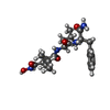  Type: peptide-like, Peptide-like / Class: Inhibitor / Mass: 448.556 Da / Num. of mol.: 1 / Source method: obtained synthetically / Formula: C23H36N4O5 Type: peptide-like, Peptide-like / Class: Inhibitor / Mass: 448.556 Da / Num. of mol.: 1 / Source method: obtained synthetically / Formula: C23H36N4O5References: N-{(2S)-2-[2-(hydroxyamino)-2-oxoethyl]-4-methylpentanoyl}-L-leucyl-L-phenylalaninamide |
-Experimental details
-Experiment
| Experiment | Method: SOLUTION NMR | ||||||||||||||||||||||||||||||||||||||||||||||||||||||||||||||||||||||||||||||||
|---|---|---|---|---|---|---|---|---|---|---|---|---|---|---|---|---|---|---|---|---|---|---|---|---|---|---|---|---|---|---|---|---|---|---|---|---|---|---|---|---|---|---|---|---|---|---|---|---|---|---|---|---|---|---|---|---|---|---|---|---|---|---|---|---|---|---|---|---|---|---|---|---|---|---|---|---|---|---|---|---|---|
| NMR experiment |
|
 HSQC
HSQC- Sample preparation
| Details |
| ||||||||||||||||||||||||||||||||||||||||||||||||||||||||||||||||||||||||||||||
|---|---|---|---|---|---|---|---|---|---|---|---|---|---|---|---|---|---|---|---|---|---|---|---|---|---|---|---|---|---|---|---|---|---|---|---|---|---|---|---|---|---|---|---|---|---|---|---|---|---|---|---|---|---|---|---|---|---|---|---|---|---|---|---|---|---|---|---|---|---|---|---|---|---|---|---|---|---|---|---|
| Sample |
| ||||||||||||||||||||||||||||||||||||||||||||||||||||||||||||||||||||||||||||||
| Sample conditions | pH: 7 / Pressure: ambient / Temperature: 305 K | ||||||||||||||||||||||||||||||||||||||||||||||||||||||||||||||||||||||||||||||
| Crystal grow | *PLUS Method: other |
-NMR measurement
| NMR spectrometer |
|
|---|
- Processing
| NMR software |
| ||||||||||||
|---|---|---|---|---|---|---|---|---|---|---|---|---|---|
| Refinement | Software ordinal: 1 Details: DISTANCE GEOMETRY (DGII INTERFACED TO INSIGHTII) FOLLOWED BY OPTIMIZATION USING SIMULATED ANNEALING WITHOUT A PHYSICAL FORCEFIELD WAS USED TO GENERATE 39 STARTING STRUCTURES. THESE 39 ...Details: DISTANCE GEOMETRY (DGII INTERFACED TO INSIGHTII) FOLLOWED BY OPTIMIZATION USING SIMULATED ANNEALING WITHOUT A PHYSICAL FORCEFIELD WAS USED TO GENERATE 39 STARTING STRUCTURES. THESE 39 STRUCTURES WERE FURTHER REFINED BY RESTRAINED MOLECULAR DYNAMICS AND RESTRAINED MINIMIZATION USING DISCOVER AND THE AMBER FORCEFIELD. THE 20 BEST STRUCTURES WITH LOWEST ENERGY AND FEWEST ABERRATIONS IN WELL-DEFINED REGIONS WERE SELECTED. RESTRAINTS INCLUDE 1336 INTERRESIDUE NOES, 55 PHI TORSION RESTRAINTS, 42 HYDROGEN BONDS, 15 METAL TO LIGAND DISTANCES, AND PEPTIDE BOND TORSION RESTRAINTS TO MAINTAIN PLANARITY. THE COMPLETE RESTRAINT LIST IS AVAILABLE AS PDB ENTRY 1UMT-MR. THE MEAN LARGEST NOE VIOLATION IS 0.64 +/- 0.07 ANGSTROM. FOR RESIDUES 83 THROUGH 250, THE MEAN BACKBONE (N, CA, C', O) RMSD TO THE AVERAGE IS 0.91 +/- 0.06 ANGSTROM. THE MEAN RMSD OF ALL HEAVY ATOMS TO THE AVERAGE IS 1.42 +/- 0.06 ANGSTROM. THIS ENSEMBLE WAS AVERAGED AND MINIMIZED WITH RESTRAINTS TO GENERATE THIS MODEL. RESIDUES 249 (167) - 256 (174) AT THE C-TERMINUS ARE DYNAMICALLY DISORDERED IN SOLUTION, JUDGING FROM THEIR LONG T2S AND LACK OF NOES. THESE RESIDUES HAVE BEEN OMITTED FROM THE MODEL. | ||||||||||||
| NMR representative | Selection criteria: minimized average structure | ||||||||||||
| NMR ensemble | Conformer selection criteria: all calculated structures submitted Conformers calculated total number: 1 / Conformers submitted total number: 1 |
