 Movie
Movie Controller


+ Open data

- Basic information
| Entry | Database: PDB / ID: 1tyu | |||||||||
|---|---|---|---|---|---|---|---|---|---|---|
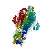
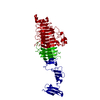
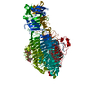
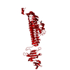
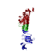
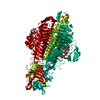
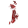
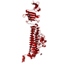
| Title | STRUCTURE OF TAILSPIKE-PROTEIN | |||||||||
 Components Components | TAILSPIKE PROTEIN | |||||||||
 Keywords Keywords | VIRAL ADHESION PROTEIN / COMPLEX / RECEPTOR / ENDOGLYCOSIDASE CARBOHYDRATE / CELL RECEPTOR / RECOGNITION / BINDING PROTEIN LIPOPOLYSACCHARIDE | |||||||||
| Function / homology |  Function and homology information Function and homology informationendo-1,3-alpha-L-rhamnosidase activity / symbiont entry into host cell via disruption of host cell envelope lipopolysaccharide / virus tail, fiber / symbiont entry into host cell via disruption of host cell envelope / symbiont entry into host / Hydrolases; Glycosylases; Glycosidases, i.e. enzymes that hydrolyse O- and S-glycosyl compounds / adhesion receptor-mediated virion attachment to host cell / virion attachment to host cell Similarity search - Function | |||||||||
| Biological species |  Enterobacteria phage P22 (virus) Enterobacteria phage P22 (virus) | |||||||||
| Method |  X-RAY DIFFRACTION / Resolution: 1.8 Å X-RAY DIFFRACTION / Resolution: 1.8 Å | |||||||||
 Authors Authors | Steinbacher, S. / Huber, R. | |||||||||
 Citation Citation | Journal: Proc.Natl.Acad.Sci.USA / Year: 1996 Title: Crystal structure of phage P22 tailspike protein complexed with Salmonella sp. O-antigen receptors. Authors: Steinbacher, S. / Baxa, U. / Miller, S. / Weintraub, A. / Seckler, R. / Huber, R. #1: Journal: Biophys.J. / Year: 1996Title: Interactions of Phage P22 Tails with Their Cellular Receptor, Salmonella O-Antigen Polysaccharide Authors: Baxa, U. / Steinbacher, S. / Miller, S. / Weintraub, A. / Huber, R. / Seckler, R. #2: Journal: Science / Year: 1994Title: Crystal Structure of P22 Tailspike Protein: Interdigitated Subunits in a Thermostable Trimer Authors: Steinbacher, S. / Seckler, R. / Miller, S. / Steipe, B. / Huber, R. / Reinemer, P. | |||||||||
| History |
|
- Structure visualization
| Structure viewer | Molecule: MolmilJmol/JSmol |
|---|
- Downloads & links
-Download
| PDBx/mmCIF format | 1tyu.cif.gz | 123.3 KB | Display | PDBx/mmCIF format |
|---|---|---|---|---|
| PDB format | pdb1tyu.ent.gz | 92.5 KB | Display | PDB format |
| PDBx/mmJSON format | 1tyu.json.gz | Tree view | PDBx/mmJSON format | |
| Others |  Other downloads Other downloads |
-Validation report
| Arichive directory | https://data.pdbj.org/pub/pdb/validation_reports/ty/1tyuftp://data.pdbj.org/pub/pdb/validation_reports/ty/1tyu | HTTPS FTP |
|---|
-Related structure data


-Links
 PDBj
PDBj- Assembly
| Deposited unit | 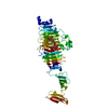
| ||||||||
|---|---|---|---|---|---|---|---|---|---|
| 1 | 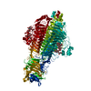
| ||||||||
| Unit cell |
|
-Components
| #1: Protein | Mass: 59616.648 Da / Num. of mol.: 1 Fragment: RESIDUES 109-666 LACKING THE N-TERMINAL, HEAD-BINDING DOMAIN Source method: isolated from a genetically manipulated source Source: (gene. exp.) Enterobacteria phage P22 (virus) / Genus: P22-like viruses / Gene: PHAGE P22 GENE 9 / Gene (production host): PHAGE P22 GENE 9 / Production host:  |
|---|---|
| #2: Polysaccharide | alpha-D-galactopyranose-(1-2)-[alpha-D-Tyvelopyranose-(1-3)]alpha-D-mannopyranose-(1-4)-alpha-L- ...alpha-D-galactopyranose-(1-2)-[alpha-D-Tyvelopyranose-(1-3)]alpha-D-mannopyranose-(1-4)-alpha-L-rhamnopyranose-(1-3)-alpha-D-galactopyranose-(1-2)-[alpha-D-Tyvelopyranose-(1-3)]alpha-D-mannopyranose-(1-4)-alpha-L-rhamnopyranose Source method: isolated from a genetically manipulated source |
| #3: Water | ChemComp-HOH /  Mass: 18.015 Da / Num. of mol.: 214 / Source method: isolated from a natural source / Formula: H2O Mass: 18.015 Da / Num. of mol.: 214 / Source method: isolated from a natural source / Formula: H2O |
-Experimental details
-Experiment
| Experiment | Method: X-RAY DIFFRACTION |
|---|
- Sample preparation
| Crystal | Density Matthews: 2.47 Å3/Da / Density % sol: 50.19 % | ||||||||||||||||||||||||||||||||||||||||||
|---|---|---|---|---|---|---|---|---|---|---|---|---|---|---|---|---|---|---|---|---|---|---|---|---|---|---|---|---|---|---|---|---|---|---|---|---|---|---|---|---|---|---|---|
| Crystal grow | pH: 7.5 Details: COMPLEX FORMED BY SOAKING WITH 2MM OCTASACCHARIDE FROM SALMONELLA ENTERITIDIS AT PH 7.5 | ||||||||||||||||||||||||||||||||||||||||||
| Crystal grow | *PLUS Temperature: 4 ℃ / pH: 10 / Method: vapor diffusion, hanging dropDetails: drop contained 0.005 ml of drop solution and 0.003 ml of precipitant | ||||||||||||||||||||||||||||||||||||||||||
| Components of the solutions | *PLUS
|
-Data collection
| Diffraction source | Wavelength: 1.5418 |
|---|---|
| Detector | Type: MARRESEARCH / Detector: IMAGE PLATE / Date: Aug 1, 1995 |
| Radiation | Monochromatic (M) / Laue (L): M / Scattering type: x-ray |
| Radiation wavelength | Wavelength: 1.5418 Å / Relative weight: 1 |
| Reflection | Num. obs: 51290 / % possible obs: 93.5 % / Redundancy: 5.4 % / Rmerge(I) obs: 0.061 |
| Reflection | *PLUS Highest resolution: 1.8 Å / Lowest resolution: 9999 Å / Num. measured all: 276996 |
| Reflection shell | *PLUS Highest resolution: 1.8 Å / Lowest resolution: 18.5 Å / % possible obs: 36.5 % / Rmerge(I) obs: 0.293 |
- Processing
| Software |
| ||||||||||||||||||||||||||||||||||||||||||||||||||||||||||||||||||||||||||||||||
|---|---|---|---|---|---|---|---|---|---|---|---|---|---|---|---|---|---|---|---|---|---|---|---|---|---|---|---|---|---|---|---|---|---|---|---|---|---|---|---|---|---|---|---|---|---|---|---|---|---|---|---|---|---|---|---|---|---|---|---|---|---|---|---|---|---|---|---|---|---|---|---|---|---|---|---|---|---|---|---|---|---|
| Refinement | Resolution: 1.8→8 Å / σ(F): 0 /
| ||||||||||||||||||||||||||||||||||||||||||||||||||||||||||||||||||||||||||||||||
| Refinement step | Cycle: LAST / Resolution: 1.8→8 Å
| ||||||||||||||||||||||||||||||||||||||||||||||||||||||||||||||||||||||||||||||||
| Refine LS restraints |
| ||||||||||||||||||||||||||||||||||||||||||||||||||||||||||||||||||||||||||||||||
| Software | *PLUS Name: X-PLOR / Classification: refinement | ||||||||||||||||||||||||||||||||||||||||||||||||||||||||||||||||||||||||||||||||
| Refinement | *PLUS | ||||||||||||||||||||||||||||||||||||||||||||||||||||||||||||||||||||||||||||||||
| Solvent computation | *PLUS | ||||||||||||||||||||||||||||||||||||||||||||||||||||||||||||||||||||||||||||||||
| Displacement parameters | *PLUS |
