 Movie
Movie Controller

[English] 日本語
 Yorodumi
- PDB-1rwt: Crystal Structure of Spinach Major Light-harvesting complex at 2.... -
+ Open data

- Basic information
| Entry | Database: PDB / ID: 1rwt | |||||||||
|---|---|---|---|---|---|---|---|---|---|---|
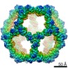
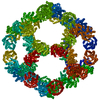
| Title | Crystal Structure of Spinach Major Light-harvesting complex at 2.72 Angstrom Resolution | |||||||||
 Components Components | Chlorophyll A-B binding protein, chloroplast | |||||||||
 Keywords Keywords | PHOTOSYNTHESIS / LIGHT-HARVESTING COMPLEX / MEMBRANE PROTEIN / PLANT | |||||||||
| Function / homology |  Function and homology information Function and homology informationphotosynthesis, light harvesting in photosystem I / photosystem I / photosystem II / chlorophyll binding / chloroplast thylakoid membrane / response to light stimulus / metal ion binding Similarity search - Function | |||||||||
| Biological species |  Spinacia oleracea (spinach) Spinacia oleracea (spinach) | |||||||||
| Method |  X-RAY DIFFRACTION / SYNCHROTRON / SIR in combination with real-space averaging refinement, extension / Resolution: 2.72 Å X-RAY DIFFRACTION / SYNCHROTRON / SIR in combination with real-space averaging refinement, extension / Resolution: 2.72 Å | |||||||||
 Authors Authors | Liu, Z. / Yan, H. / Wang, K. / Kuang, T. / Zhang, J. / Gui, L. / An, X. / Chang, W. | |||||||||
 Citation Citation | Journal: Nature / Year: 2004 Title: Crystal structure of spinach major light-harvesting complex at 2.72 A resolution Authors: Liu, Z. / Yan, H. / Wang, K. / Kuang, T. / Zhang, J. / Gui, L. / An, X. / Chang, W. | |||||||||
| History |
|
- Structure visualization
| Structure viewer | Molecule: MolmilJmol/JSmol |
|---|
- Downloads & links
-Download
| PDBx/mmCIF format | 1rwt.cif.gz | 718.3 KB | Display | PDBx/mmCIF format |
|---|---|---|---|---|
| PDB format | pdb1rwt.ent.gz | 661 KB | Display | PDB format |
| PDBx/mmJSON format | 1rwt.json.gz | Tree view | PDBx/mmJSON format | |
| Others |  Other downloads Other downloads |
-Validation report
| Arichive directory | https://data.pdbj.org/pub/pdb/validation_reports/rw/1rwtftp://data.pdbj.org/pub/pdb/validation_reports/rw/1rwt | HTTPS FTP |
|---|
-Related structure data
| Similar structure data |
|---|

-Links
 PDBj
PDBj


- Assembly
| Deposited unit | 
| ||||||||
|---|---|---|---|---|---|---|---|---|---|
| 1 | x 6
| ||||||||
| Unit cell |
| ||||||||
| Components on special symmetry positions |
|
-Components
-Protein , 1 types, 10 molecules ABCDEFGHIJ
| #1: Protein | Mass: 25000.330 Da / Num. of mol.: 10 / Source method: isolated from a natural source / Source: (natural) Spinacia oleracea (spinach) / Tissue: leaf / References: UniProt: P12333 |
|---|
-Sugars , 2 types, 20 molecules 

| #2: Sugar | ChemComp-BNG /  Type: D-saccharide / Mass: 306.395 Da / Num. of mol.: 10 Type: D-saccharide / Mass: 306.395 Da / Num. of mol.: 10Source method: isolated from a genetically manipulated source Formula: C15H30O6 / Comment: detergent*YM #8: Sugar | ChemComp-DGD /  Type: saccharide / Mass: 949.299 Da / Num. of mol.: 10 / Source method: obtained synthetically / Formula: C51H96O15 Type: saccharide / Mass: 949.299 Da / Num. of mol.: 10 / Source method: obtained synthetically / Formula: C51H96O15 |
|---|
-Non-polymers , 8 types, 890 molecules 


| #3: Chemical | ChemComp-NA /  Mass: 22.990 Da / Num. of mol.: 1 / Source method: obtained synthetically / Formula: Na Mass: 22.990 Da / Num. of mol.: 1 / Source method: obtained synthetically / Formula: Na | ||||||||||||
|---|---|---|---|---|---|---|---|---|---|---|---|---|---|
| #4: Chemical | ChemComp-LUT / (  Mass: 568.871 Da / Num. of mol.: 20 / Source method: obtained synthetically / Formula: C40H56O2 Mass: 568.871 Da / Num. of mol.: 20 / Source method: obtained synthetically / Formula: C40H56O2#5: Chemical | ChemComp-XAT / (  Mass: 600.870 Da / Num. of mol.: 10 / Source method: obtained synthetically / Formula: C40H56O4 Mass: 600.870 Da / Num. of mol.: 10 / Source method: obtained synthetically / Formula: C40H56O4#6: Chemical | ChemComp-NEX / (  Mass: 600.870 Da / Num. of mol.: 10 / Source method: obtained synthetically / Formula: C40H56O4 Mass: 600.870 Da / Num. of mol.: 10 / Source method: obtained synthetically / Formula: C40H56O4#7: Chemical | ChemComp-LHG /  Mass: 722.970 Da / Num. of mol.: 10 / Source method: obtained synthetically / Formula: C38H75O10P / Comment: phospholipid*YM Mass: 722.970 Da / Num. of mol.: 10 / Source method: obtained synthetically / Formula: C38H75O10P / Comment: phospholipid*YM#9: Chemical | ChemComp-CHL /  Mass: 907.472 Da / Num. of mol.: 60 / Source method: obtained synthetically / Formula: C55H70MgN4O6 Mass: 907.472 Da / Num. of mol.: 60 / Source method: obtained synthetically / Formula: C55H70MgN4O6#10: Chemical | ChemComp-CLA /  Mass: 893.489 Da / Num. of mol.: 80 / Source method: obtained synthetically / Formula: C55H72MgN4O5 Mass: 893.489 Da / Num. of mol.: 80 / Source method: obtained synthetically / Formula: C55H72MgN4O5#11: Water | ChemComp-HOH / | Mass: 18.015 Da / Num. of mol.: 699 / Source method: isolated from a natural source / Formula: H2O |
-Experimental details
-Experiment
| Experiment | Method: X-RAY DIFFRACTION / Number of used crystals: 1 |
|---|
- Sample preparation
| Crystal | Density Matthews: 5.36 Å3/Da / Density % sol: 76.86 % |
|---|---|
| Crystal grow | Temperature: 291 K / Method: vapor diffusion / pH: 7.5 Details: Citrate trisodium, beta-nonyl-glucoside, deoxy-bigchap, DGDG, Hepes, pH 7.5, VAPOR DIFFUSION, temperature 291K |
-Data collection
| Diffraction |
| |||||||||||||||
|---|---|---|---|---|---|---|---|---|---|---|---|---|---|---|---|---|
| Diffraction source |
| |||||||||||||||
| Detector |
| |||||||||||||||
| Radiation | Protocol: SINGLE WAVELENGTH / Monochromatic (M) / Laue (L): M / Scattering type: x-ray | |||||||||||||||
| Radiation wavelength | Wavelength: 1 Å / Relative weight: 1 | |||||||||||||||
| Reflection | Resolution: 2.7→25 Å / Num. all: 211079 / Num. obs: 191660 / % possible obs: 90.8 % / Observed criterion σ(I): -3 / Redundancy: 5.2 % / Biso Wilson estimate: 20.5 Å2 / Rmerge(I) obs: 0.082 / Net I/σ(I): 14.4 | |||||||||||||||
| Reflection shell | Resolution: 2.7→2.75 Å / Rmerge(I) obs: 0.368 / Mean I/σ(I) obs: 2.5 / Num. unique all: 9188 / % possible all: 79.6 |


- Processing
| Software |
| |||||||||||||||||||||||||
|---|---|---|---|---|---|---|---|---|---|---|---|---|---|---|---|---|---|---|---|---|---|---|---|---|---|---|
| Refinement | Method to determine structure: SIR in combination with real-space averaging refinement, extension Resolution: 2.72→10 Å / Rfactor Rfree error: 0.002 / Data cutoff high absF: 557510.7 / Data cutoff low absF: 0 / Isotropic thermal model: RESTRAINED / Cross valid method: THROUGHOUT / σ(F): 0 / Stereochemistry target values: Engh & Huber
| |||||||||||||||||||||||||
| Solvent computation | Solvent model: FLAT MODEL / Bsol: 54.7972 Å2 / ksol: 0.37093 e/Å3 | |||||||||||||||||||||||||
| Displacement parameters | Biso mean: 31.3 Å2
| |||||||||||||||||||||||||
| Refine analyze |
| |||||||||||||||||||||||||
| Refinement step | Cycle: LAST / Resolution: 2.72→10 Å
| |||||||||||||||||||||||||
| Refine LS restraints |
| |||||||||||||||||||||||||
| LS refinement shell | Resolution: 2.72→2.89 Å / Rfactor Rfree error: 0.007 / Total num. of bins used: 6
| |||||||||||||||||||||||||
| Xplor file |
|
