 Movie
Movie Controller

[English] 日本語
 Yorodumi
- PDB-1qiv: CALMODULIN COMPLEXED WITH N-(3,3,-DIPHENYLPROPYL)-N'-[1-R-(3,4-BI... -
+ Open data

- Basic information
| Entry | Database: PDB / ID: 1qiv | ||||||
|---|---|---|---|---|---|---|---|
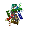
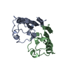
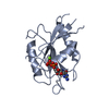
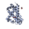
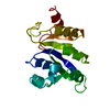
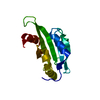
| Title | CALMODULIN COMPLEXED WITH N-(3,3,-DIPHENYLPROPYL)-N'-[1-R-(3,4-BIS-BUTOXYPHENYL)-ETHYL]-PROPYLENEDIAMINE (DPD), 1:2 COMPLEX | ||||||
 Components Components | CALMODULIN | ||||||
 Keywords Keywords | CALCIUM-BINDING PROTEIN | ||||||
| Function / homology |  Function and homology information Function and homology informationregulation of store-operated calcium channel activity / : / : / regulation of response to tumor cell / positive regulation of autophagic cell death / DAPK1-calmodulin complex / : / : / : / : ...regulation of store-operated calcium channel activity / : / : / regulation of response to tumor cell / positive regulation of autophagic cell death / DAPK1-calmodulin complex / : / : / : / : / : / establishment of protein localization to mitochondrial membrane / type 3 metabotropic glutamate receptor binding / establishment of protein localization to membrane / positive regulation of DNA binding / negative regulation of high voltage-gated calcium channel activity / negative regulation of ryanodine-sensitive calcium-release channel activity / organelle localization by membrane tethering / mitochondrion-endoplasmic reticulum membrane tethering / autophagosome membrane docking / negative regulation of calcium ion export across plasma membrane / regulation of cardiac muscle cell action potential / nitric-oxide synthase binding / regulation of synaptic vesicle exocytosis / adenylate cyclase binding / regulation of ryanodine-sensitive calcium-release channel activity / protein phosphatase activator activity / catalytic complex / detection of calcium ion / regulation of synaptic vesicle endocytosis / regulation of cardiac muscle contraction / activation of adenylate cyclase activity / phosphatidylinositol 3-kinase binding / calcium channel inhibitor activity / positive regulation of nitric-oxide synthase activity / regulation of release of sequestered calcium ion into cytosol by sarcoplasmic reticulum / enzyme regulator activity / titin binding / regulation of calcium-mediated signaling / voltage-gated potassium channel complex / potassium ion transmembrane transport / calcium channel complex / regulation of heart rate / response to amphetamine / adenylate cyclase activator activity / sarcomere / nitric-oxide synthase regulator activity / regulation of cytokinesis / spindle microtubule / calcium channel regulator activity / calcium-mediated signaling / response to calcium ion / G2/M transition of mitotic cell cycle / spindle pole / disordered domain specific binding / calcium-dependent protein binding / myelin sheath / protein autophosphorylation / growth cone / vesicle / transmembrane transporter binding / neuron projection / positive regulation of apoptotic process / protein domain specific binding / calcium ion binding / centrosome / protein kinase binding / protein-containing complex / mitochondrion / nucleoplasm / nucleus / plasma membrane / cytoplasm / cytosol Similarity search - Function | ||||||
| Biological species |  | ||||||
| Method |  X-RAY DIFFRACTION / MOLECULAR REPLACEMENT / Resolution: 2.64 Å X-RAY DIFFRACTION / MOLECULAR REPLACEMENT / Resolution: 2.64 Å | ||||||
 Authors Authors | Harmat, V. / Bocskei, Z.S. / Vertessy, B.G. / Naray-Szabo, G. / Ovadi, J. | ||||||
 Citation Citation | Journal: J.Mol.Biol. / Year: 2000 Title: A New Potent Calmodulin Antagonist with Arylalkylamine Structure: Crystallographic, Spectroscopic and Functional Studies Authors: Harmat, V. / Bocskei, Z.S. / Naray-Szabo, G. / Bata, I. / Csutor, A.S. / Hermecz, I. / Aranyi, P. / Szabo, B. / Liliom, K. / Vertessy, B.G. / Ovadi, J. #1: Journal: Biochemistry / Year: 1998Title: Simultaneous Binding of Drugs with Different Chemical Structures to Ca 2+ Calmodulin: Crystallographic and Spectroscopic Studies Authors: Vertessy, B.G. / Harmat, V. / Bocskei, Z.S. / Naray-Szabo, G. / Orosz, F. / Ovadi, J. #2: Journal: Proteins: Struct.,Funct., Genet. / Year: 1997 Title: Crystallization and Preliminary Diffraction Analysis of Ca(2+)-Calmodulin-Drug and Apocalmodulin-Drug Complexes. Authors: Vertessy, B.G. / Bocskei, Z.S. / Harmath, V. / Naray-Szabo, G. / Ovadi, J. #3: Journal: Nat.Struct.Biol. / Year: 1994Title: Trifluoperazine-Induced Conformational Change in Ca (2+)-Calmodulin Authors: Vandonselaar, M. / Hickie, R.A. / Quail, J.W. / Delbaere, L.T.J. #4: Journal: Biochemistry / Year: 1994Title: Drug Binding by Calmodulin: Crystal Structure of a Calmodulin-Trifluoperazine Complex Authors: Cook, W.J. / Walter, L.J. / Walter, M.R. | ||||||
| History |
|
- Structure visualization
| Structure viewer | Molecule: MolmilJmol/JSmol |
|---|
- Downloads & links
-Download
| PDBx/mmCIF format | 1qiv.cif.gz | 45.7 KB | Display | PDBx/mmCIF format |
|---|---|---|---|---|
| PDB format | pdb1qiv.ent.gz | 30.9 KB | Display | PDB format |
| PDBx/mmJSON format | 1qiv.json.gz | Tree view | PDBx/mmJSON format | |
| Others |  Other downloads Other downloads |
-Validation report
| Summary document | 1qiv_validation.pdf.gz | 1.1 MB | Display | wwPDB validaton report |
|---|---|---|---|---|
| Full document | 1qiv_full_validation.pdf.gz | 1.1 MB | Display | |
| Data in XML | 1qiv_validation.xml.gz | 9.2 KB | Display | |
| Data in CIF | 1qiv_validation.cif.gz | 11 KB | Display | |
| Arichive directory | https://data.pdbj.org/pub/pdb/validation_reports/qi/1qivftp://data.pdbj.org/pub/pdb/validation_reports/qi/1qiv | HTTPS FTP |
-Related structure data
| Related structure data | 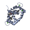 1qiwC 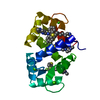 1linS S: Starting model for refinement C: citing same article ( |
|---|---|
| Similar structure data |


-Links
 PDBj
PDBj

- Assembly
| Deposited unit | 
| ||||||||
|---|---|---|---|---|---|---|---|---|---|
| 1 |
| ||||||||
| Unit cell |
|
-Components
| #1: Protein | Mass: 16721.350 Da / Num. of mol.: 1 / Source method: isolated from a natural source / Source: (natural) | ||
|---|---|---|---|
| #2: Chemical | ChemComp-CA /   Mass: 40.078 Da / Num. of mol.: 4 / Source method: obtained synthetically / Formula: Ca Mass: 40.078 Da / Num. of mol.: 4 / Source method: obtained synthetically / Formula: Ca#3: Chemical |   Mass: 516.757 Da / Num. of mol.: 2 / Source method: obtained synthetically / Formula: C34H48N2O2 Mass: 516.757 Da / Num. of mol.: 2 / Source method: obtained synthetically / Formula: C34H48N2O2 |
-Experimental details
-Experiment
| Experiment | Method: X-RAY DIFFRACTION / Number of used crystals: 1 |
|---|
- Sample preparation
| Crystal | Density Matthews: 2.5 Å3/Da / Density % sol: 52 % | ||||||||||||||||||||||||||||||||||||||||||||||||||||||
|---|---|---|---|---|---|---|---|---|---|---|---|---|---|---|---|---|---|---|---|---|---|---|---|---|---|---|---|---|---|---|---|---|---|---|---|---|---|---|---|---|---|---|---|---|---|---|---|---|---|---|---|---|---|---|---|
| Crystal grow | Method: vapor diffusion, hanging drop / pH: 5 Details: PROTEIN WAS CRYSTALLIZED BY HANGING DROP TECHNIQUE AT ROOM TEMPERATURE FROM 50 MM PH=5.0 SODIUM CACODYLATE/HCL BUFFER, 10 MM MGCL2, 10 MM CACL2, 2MM DPD AND 28% PEG 8000. CRYSTAL GROWTH TOOK 2-3 WEEKS., pH 5.00 | ||||||||||||||||||||||||||||||||||||||||||||||||||||||
| Crystal | *PLUS | ||||||||||||||||||||||||||||||||||||||||||||||||||||||
| Crystal grow | *PLUS pH: 7 / Method: vapor diffusion, hanging drop | ||||||||||||||||||||||||||||||||||||||||||||||||||||||
| Components of the solutions | *PLUS
|
-Data collection
| Diffraction | Mean temperature: 93 K |
|---|---|
| Diffraction source | Source: ROTATING ANODE / Type: RIGAKU RUH2R / Wavelength: 1.5418 |
| Detector | Type: MARRESEARCH / Detector: IMAGE PLATE / Date: Aug 15, 1997 / Details: NORMAL FOCUS |
| Radiation | Monochromator: GRAPHITE(002) / Protocol: SINGLE WAVELENGTH / Monochromatic (M) / Laue (L): M / Scattering type: x-ray |
| Radiation wavelength | Wavelength: 1.5418 Å / Relative weight: 1 |
| Reflection | Resolution: 2.63→24.58 Å / Num. obs: 4969 / % possible obs: 92.7 % / Observed criterion σ(I): 0 / Redundancy: 4.3 % / Biso Wilson estimate: 28.4 Å2 / Rmerge(I) obs: 0.058 / Net I/σ(I): 20.6 |
| Reflection shell | Resolution: 2.63→2.72 Å / Redundancy: 2.3 % / Rmerge(I) obs: 0.123 / Mean I/σ(I) obs: 9 / % possible all: 76.2 |
| Reflection | *PLUS Num. measured all: 107694 |
- Processing
| Software |
| ||||||||||||||||||||||||||||||||||||||||||||||||||||||||||||
|---|---|---|---|---|---|---|---|---|---|---|---|---|---|---|---|---|---|---|---|---|---|---|---|---|---|---|---|---|---|---|---|---|---|---|---|---|---|---|---|---|---|---|---|---|---|---|---|---|---|---|---|---|---|---|---|---|---|---|---|---|---|
| Refinement | Method to determine structure: MOLECULAR REPLACEMENT Starting model: PDB ENTRY 1LIN Resolution: 2.64→24.58 Å / Rfactor Rfree error: 0.01 / Data cutoff high absF: 1000000 / Data cutoff low absF: 0.001 / Isotropic thermal model: GROUP / Cross valid method: THROUGHOUT / σ(F): 0 Details: DISORDERED SIDE CHAIN ATOMS OF RESIDUES ARG 74 - THR 79 AND SER 81 OF THE CENTRAL REGION, GLU 6, GLU 83, TRIMETHYL-LYSINE 115, GLU 123 AND GLU 127 ARE NOT SEEN IN THE CRYSTAL STRUCTURE, AS ...Details: DISORDERED SIDE CHAIN ATOMS OF RESIDUES ARG 74 - THR 79 AND SER 81 OF THE CENTRAL REGION, GLU 6, GLU 83, TRIMETHYL-LYSINE 115, GLU 123 AND GLU 127 ARE NOT SEEN IN THE CRYSTAL STRUCTURE, AS WELL AS THE N-TERMINAL ALA 1, ASP 2 AND C-TERMINAL ALA 147, LYS 148 RESIDUES. TEMPERATURE FACTORS FOR THE ATOMS IN RESIDUES 75 - 81 ARE UNUSUALLY HIGH, INDICATING FLEXIBILITY IN THIS REGION. THE C-TERMINAL RESIDUES WERE NOT SEEN IN THE DENSITY MAPS
| ||||||||||||||||||||||||||||||||||||||||||||||||||||||||||||
| Displacement parameters | Biso mean: 28 Å2
| ||||||||||||||||||||||||||||||||||||||||||||||||||||||||||||
| Refine analyze |
| ||||||||||||||||||||||||||||||||||||||||||||||||||||||||||||
| Refinement step | Cycle: LAST / Resolution: 2.64→24.58 Å
| ||||||||||||||||||||||||||||||||||||||||||||||||||||||||||||
| Refine LS restraints |
| ||||||||||||||||||||||||||||||||||||||||||||||||||||||||||||
| LS refinement shell | Resolution: 2.64→2.76 Å / Rfactor Rfree error: 0.085 / Total num. of bins used: 8
| ||||||||||||||||||||||||||||||||||||||||||||||||||||||||||||
| Xplor file |
| ||||||||||||||||||||||||||||||||||||||||||||||||||||||||||||
| Software | *PLUS Name: X-PLOR / Version: 3.851 / Classification: refinement | ||||||||||||||||||||||||||||||||||||||||||||||||||||||||||||
| Refine LS restraints | *PLUS
|
