 Movie
Movie Controller

[English] 日本語
 Yorodumi
- PDB-1nfs: STRUCTURE AND MECHANISM OF ACTION OF ISOPENTENYLPYROPHOSPHATE-DIM... -
+ Open data

- Basic information
| Entry | Database: PDB / ID: 1nfs | ||||||
|---|---|---|---|---|---|---|---|
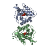
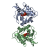
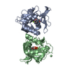
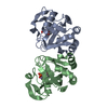
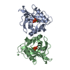
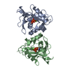
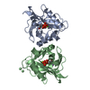
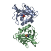
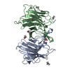
| Title | STRUCTURE AND MECHANISM OF ACTION OF ISOPENTENYLPYROPHOSPHATE-DIMETHYLALLYLPYROPHOSPHATE ISOMERASE: COMPLEX WITH NIPP | ||||||
 Components Components | ISOPENTENYL-DIPHOSPHATE DELTA-ISOMERASE | ||||||
 Keywords Keywords | ISOMERASE / COMPLEX | ||||||
| Function / homology |  Function and homology information Function and homology informationisopentenyl-diphosphate Delta-isomerase / isopentenyl-diphosphate delta-isomerase activity / dimethylallyl diphosphate biosynthetic process / isoprenoid biosynthetic process / DNA damage response / magnesium ion binding / zinc ion binding / cytoplasm Similarity search - Function | ||||||
| Biological species |  | ||||||
| Method |  X-RAY DIFFRACTION / MOLECULAR REPLACEMENT / Resolution: 1.96 Å X-RAY DIFFRACTION / MOLECULAR REPLACEMENT / Resolution: 1.96 Å | ||||||
 Authors Authors | Wouters, J. | ||||||
 Citation Citation | Journal: J.Biol.Chem. / Year: 2003 Title: Catalytic Mechanism of Escherichia coli Isopentenyl Diphosphate Isomerase Involves Cys-67, Glu-116, and Tyr-104 as Suggested by Crystal Structures of Complexes with Transition State Analogues ...Title: Catalytic Mechanism of Escherichia coli Isopentenyl Diphosphate Isomerase Involves Cys-67, Glu-116, and Tyr-104 as Suggested by Crystal Structures of Complexes with Transition State Analogues and Irreversible Inhibitors Authors: Wouters, J. / Oudjama, Y. / Barkley, S.J. / Tricot, C. / Stalon, V. / Droogmans, L. / Poulter, C.D. | ||||||
| History |
|
- Structure visualization
| Structure viewer | Molecule: MolmilJmol/JSmol |
|---|
- Downloads & links
-Download
| PDBx/mmCIF format | 1nfs.cif.gz | 87.4 KB | Display | PDBx/mmCIF format |
|---|---|---|---|---|
| PDB format | pdb1nfs.ent.gz | 64.6 KB | Display | PDB format |
| PDBx/mmJSON format | 1nfs.json.gz | Tree view | PDBx/mmJSON format | |
| Others |  Other downloads Other downloads |
-Validation report
| Arichive directory | https://data.pdbj.org/pub/pdb/validation_reports/nf/1nfsftp://data.pdbj.org/pub/pdb/validation_reports/nf/1nfs | HTTPS FTP |
|---|
-Related structure data
| Related structure data |  1nfzC  1hztS C: citing same article ( S: Starting model for refinement |
|---|---|
| Similar structure data |

-Links
 PDBj
PDBj

- Assembly
| Deposited unit | 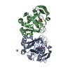
| ||||||||
|---|---|---|---|---|---|---|---|---|---|
| 1 |
| ||||||||
| Unit cell |
| ||||||||
| Details | Two molecules in the asymertic unit form the biological assembly |
-Components
| #1: Protein | Mass: 20644.391 Da / Num. of mol.: 2 Source method: isolated from a genetically manipulated source Source: (gene. exp.) References: UniProt: Q46822, isopentenyl-diphosphate Delta-isomerase #2: Chemical |   Mass: 54.938 Da / Num. of mol.: 2 / Source method: obtained synthetically / Formula: Mn Mass: 54.938 Da / Num. of mol.: 2 / Source method: obtained synthetically / Formula: Mn#3: Chemical |   Mass: 24.305 Da / Num. of mol.: 2 / Source method: obtained synthetically / Formula: Mg Mass: 24.305 Da / Num. of mol.: 2 / Source method: obtained synthetically / Formula: Mg#4: Chemical |   Mass: 249.096 Da / Num. of mol.: 2 / Source method: obtained synthetically / Formula: C4H13NO7P2 Mass: 249.096 Da / Num. of mol.: 2 / Source method: obtained synthetically / Formula: C4H13NO7P2#5: Water | ChemComp-HOH / |  Mass: 18.015 Da / Num. of mol.: 117 / Source method: isolated from a natural source / Formula: H2O Mass: 18.015 Da / Num. of mol.: 117 / Source method: isolated from a natural source / Formula: H2O |
|---|
-Experimental details
-Experiment
| Experiment | Method: X-RAY DIFFRACTION / Number of used crystals: 1 |
|---|
- Sample preparation
| Crystal | Density Matthews: 2.43 Å3/Da / Density % sol: 54.28 % | ||||||||||||||||||||||||||||||||||||||||||
|---|---|---|---|---|---|---|---|---|---|---|---|---|---|---|---|---|---|---|---|---|---|---|---|---|---|---|---|---|---|---|---|---|---|---|---|---|---|---|---|---|---|---|---|
| Crystal grow | Temperature: 290 K / Method: vapor diffusion, hanging drop / pH: 5.5 Details: PEG 2000, MANGANESE CHLORIDE, pH 5.5, VAPOR DIFFUSION, HANGING DROP, temperature 290K | ||||||||||||||||||||||||||||||||||||||||||
| Crystal grow | *PLUS Method: vapor diffusion, hanging drop | ||||||||||||||||||||||||||||||||||||||||||
| Components of the solutions | *PLUS
|
-Data collection
| Diffraction | Mean temperature: 100 K |
|---|---|
| Diffraction source | Source: ROTATING ANODE / Type: ENRAF-NONIUS / Wavelength: 1.54179 / Wavelength: 1.54179 Å |
| Detector | Type: MARRESEARCH / Detector: IMAGE PLATE / Details: MIRRORS |
| Radiation | Monochromator: graphite / Protocol: SINGLE WAVELENGTH / Monochromatic (M) / Laue (L): M / Scattering type: x-ray |
| Radiation wavelength | Wavelength: 1.54179 Å / Relative weight: 1 |
| Reflection | Resolution: 1.96→10 Å / Num. all: 27788 / Num. obs: 23302 / % possible obs: 94.8 % / Observed criterion σ(F): 2 / Observed criterion σ(I): 4 / Rmerge(I) obs: 0.042 / Net I/σ(I): 9.2 |
| Reflection shell | Resolution: 1.97→2.08 Å / Rmerge(I) obs: 0.244 / Mean I/σ(I) obs: 6.3 / % possible all: 94.8 |
| Reflection | *PLUS Highest resolution: 1.97 Å / Num. measured all: 107068 |
| Reflection shell | *PLUS % possible obs: 94.8 % |
- Processing
| Software |
| |||||||||||||||||||||||||||||||||
|---|---|---|---|---|---|---|---|---|---|---|---|---|---|---|---|---|---|---|---|---|---|---|---|---|---|---|---|---|---|---|---|---|---|---|
| Refinement | Method to determine structure: MOLECULAR REPLACEMENT Starting model: PDB ENTRY 1HZT Resolution: 1.96→10 Å / Num. parameters: 11935 / Num. restraintsaints: 15429 / Cross valid method: THROUGHOUT / σ(F): 0 / Stereochemistry target values: ENGH AND HUBER
| |||||||||||||||||||||||||||||||||
| Refine analyze | Num. disordered residues: 0 / Occupancy sum hydrogen: 0 / Occupancy sum non hydrogen: 2983 | |||||||||||||||||||||||||||||||||
| Refinement step | Cycle: LAST / Resolution: 1.96→10 Å
| |||||||||||||||||||||||||||||||||
| Refine LS restraints |
| |||||||||||||||||||||||||||||||||
| Software | *PLUS Name: SHELXL / Version: 97 / Classification: refinement | |||||||||||||||||||||||||||||||||
| Refinement | *PLUS Highest resolution: 1.97 Å / Lowest resolution: 10 Å / % reflection Rfree: 10 % / Rfactor all: 0.222 / Rfactor Rfree: 0.262 / Rfactor Rwork: 0.21 | |||||||||||||||||||||||||||||||||
| Solvent computation | *PLUS | |||||||||||||||||||||||||||||||||
| Displacement parameters | *PLUS | |||||||||||||||||||||||||||||||||
| Refine LS restraints | *PLUS
|
