 Movie
Movie Controller

[English] 日本語
 Yorodumi
- PDB-1naa: Cellobiose Dehydrogenase Flavoprotein Fragment in Complex with Ce... -
+ Open data

- Basic information
| Entry | Database: PDB / ID: 1naa | |||||||||
|---|---|---|---|---|---|---|---|---|---|---|
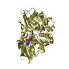
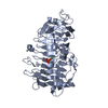
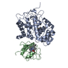
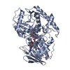
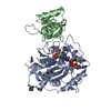
| Title | Cellobiose Dehydrogenase Flavoprotein Fragment in Complex with Cellobionolactam | |||||||||
 Components Components | Cellobiose dehydrogenase | |||||||||
 Keywords Keywords | OXIDOREDUCTASE / GMC oxidoreductase / alpha/beta structure / rossmann fold / PHBH fold / product analogue complex / 6-hydroxylated FAD | |||||||||
| Function / homology |  Function and homology information Function and homology informationcellobiose dehydrogenase (acceptor) / cellobiose dehydrogenase (acceptor) activity / cellulose catabolic process / flavin adenine dinucleotide binding / extracellular region / metal ion binding Similarity search - Function | |||||||||
| Biological species |  Phanerochaete chrysosporium (fungus) Phanerochaete chrysosporium (fungus) | |||||||||
| Method |  X-RAY DIFFRACTION / SYNCHROTRON / FOURIER SYNTHESIS / Resolution: 1.8 Å X-RAY DIFFRACTION / SYNCHROTRON / FOURIER SYNTHESIS / Resolution: 1.8 Å | |||||||||
 Authors Authors | Hallberg, B.M. / Henriksson, G. / Pettersson, G. / Vasella, A. / Divne, C. | |||||||||
 Citation Citation | Journal: J.BIOL.CHEM. / Year: 2003 Title: Mechanism of the reductive half-reaction in cellobiose dehydrogenase Authors: Hallberg, B.M. / Henriksson, G. / Pettersson, G. / Vasella, A. / Divne, C. #1: Journal: J.Mol.Biol. / Year: 2002Title: Crystal structure of the flavoprotein domain of the extracellular flavocytochrome cellobiose dehydrogenase Authors: Hallberg, B.M. / Henriksson, G. / Pettersson, G. / Divne, C. | |||||||||
| History |
|
- Structure visualization
| Structure viewer | Molecule: MolmilJmol/JSmol |
|---|
- Downloads & links
-Download
| PDBx/mmCIF format | 1naa.cif.gz | 237.8 KB | Display | PDBx/mmCIF format |
|---|---|---|---|---|
| PDB format | pdb1naa.ent.gz | 187.1 KB | Display | PDB format |
| PDBx/mmJSON format | 1naa.json.gz | Tree view | PDBx/mmJSON format | |
| Others |  Other downloads Other downloads |
-Validation report
| Summary document | 1naa_validation.pdf.gz | 1.6 MB | Display | wwPDB validaton report |
|---|---|---|---|---|
| Full document | 1naa_full_validation.pdf.gz | 1.6 MB | Display | |
| Data in XML | 1naa_validation.xml.gz | 49.7 KB | Display | |
| Data in CIF | 1naa_validation.cif.gz | 75.3 KB | Display | |
| Arichive directory | https://data.pdbj.org/pub/pdb/validation_reports/na/1naaftp://data.pdbj.org/pub/pdb/validation_reports/na/1naa | HTTPS FTP |
-Related structure data
| Related structure data |  1kdgS S: Starting model for refinement |
|---|---|
| Similar structure data |


-Links
 PDBj
PDBj
- Assembly
| Deposited unit | 
| ||||||||
|---|---|---|---|---|---|---|---|---|---|
| 1 | 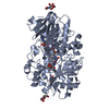
| ||||||||
| 2 | 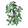
| ||||||||
| Unit cell |
|
-Components
| #1: Protein | Mass: 57544.973 Da / Num. of mol.: 2 / Fragment: c-terminal flavoprotein fragment / Source method: isolated from a natural source / Source: (natural) Phanerochaete chrysosporium (fungus) / Strain: K3References: UniProt: Q01738, cellobiose dehydrogenase (acceptor) #2: Sugar | ChemComp-NAG /   Type: D-saccharide, beta linking / Mass: 221.208 Da / Num. of mol.: 5 Type: D-saccharide, beta linking / Mass: 221.208 Da / Num. of mol.: 5Source method: isolated from a genetically manipulated source Formula: C8H15NO6 #3: Chemical |   Mass: 801.549 Da / Num. of mol.: 2 / Source method: obtained synthetically / Formula: C27H33N9O16P2 Mass: 801.549 Da / Num. of mol.: 2 / Source method: obtained synthetically / Formula: C27H33N9O16P2#4: Sugar |   Type: D-saccharide / Mass: 339.296 Da / Num. of mol.: 2 / Source method: obtained synthetically / Formula: C12H21NO10 Type: D-saccharide / Mass: 339.296 Da / Num. of mol.: 2 / Source method: obtained synthetically / Formula: C12H21NO10#5: Water | ChemComp-HOH / |  Mass: 18.015 Da / Num. of mol.: 1007 / Source method: isolated from a natural source / Formula: H2O Mass: 18.015 Da / Num. of mol.: 1007 / Source method: isolated from a natural source / Formula: H2OHas protein modification | Y | |
|---|
-Experimental details
-Experiment
| Experiment | Method: X-RAY DIFFRACTION / Number of used crystals: 1 |
|---|
- Sample preparation
| Crystal | Density Matthews: 2.07 Å3/Da / Density % sol: 40.02 % | ||||||||||||||||||||||||||||||
|---|---|---|---|---|---|---|---|---|---|---|---|---|---|---|---|---|---|---|---|---|---|---|---|---|---|---|---|---|---|---|---|
| Crystal grow | Temperature: 288 K / Method: vapor diffusion, hanging drop / pH: 6.5 Details: ammonium sulfate, dioxane, mes, cellobionolactam, pH 6.5, VAPOR DIFFUSION, HANGING DROP, temperature 288K | ||||||||||||||||||||||||||||||
| Crystal grow | *PLUS Temperature: 15 ℃ / Details: Hallberg, B.M., (2002) J.Mol.Biol., 315, 421. | ||||||||||||||||||||||||||||||
| Components of the solutions | *PLUS
|
-Data collection
| Diffraction | Mean temperature: 100 K |
|---|---|
| Diffraction source | Source: SYNCHROTRON / Site: ESRF  / Beamline: ID14-4 / Wavelength: 0.977 Å / Beamline: ID14-4 / Wavelength: 0.977 Å |
| Detector | Type: ADSC QUANTUM 4 / Detector: CCD / Date: Feb 6, 1999 |
| Radiation | Protocol: SINGLE WAVELENGTH / Monochromatic (M) / Laue (L): M / Scattering type: x-ray |
| Radiation wavelength | Wavelength: 0.977 Å / Relative weight: 1 |
| Reflection | Resolution: 1.8→57 Å / Num. all: 98043 / Num. obs: 95043 / % possible obs: 97.6 % / Observed criterion σ(F): 0 / Observed criterion σ(I): 0 / Redundancy: 4 % / Rmerge(I) obs: 0.089 / Net I/σ(I): 6.7 |
| Reflection shell | Resolution: 1.8→1.9 Å / Redundancy: 2.2 % / Rmerge(I) obs: 0.498 / Mean I/σ(I) obs: 1.5 / % possible all: 85.4 |
| Reflection | *PLUS Lowest resolution: 57 Å / Num. measured all: 376799 |
| Reflection shell | *PLUS % possible obs: 85.4 % |
- Processing
| Software |
| |||||||||||||||||||||
|---|---|---|---|---|---|---|---|---|---|---|---|---|---|---|---|---|---|---|---|---|---|---|
| Refinement | Method to determine structure: FOURIER SYNTHESIS Starting model: PDB ENTRY 1KDG Resolution: 1.8→30 Å / σ(F): 0 / Stereochemistry target values: Engh & Huber
| |||||||||||||||||||||
| Refinement step | Cycle: LAST / Resolution: 1.8→30 Å
| |||||||||||||||||||||
| Refine LS restraints |
| |||||||||||||||||||||
| Refinement | *PLUS Lowest resolution: 30 Å | |||||||||||||||||||||
| Solvent computation | *PLUS | |||||||||||||||||||||
| Displacement parameters | *PLUS | |||||||||||||||||||||
| Refine LS restraints | *PLUS
|
