 Movie
Movie Controller

[English] 日本語
 Yorodumi
- PDB-1m4h: Crystal Structure of Beta-secretase complexed with Inhibitor OM00-3 -
+ Open data

- Basic information
| Entry | Database: PDB / ID: 1m4h | |||||||||
|---|---|---|---|---|---|---|---|---|---|---|
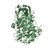
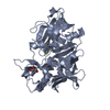
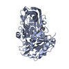
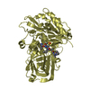
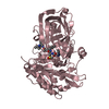
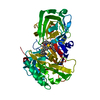
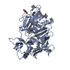
| Title | Crystal Structure of Beta-secretase complexed with Inhibitor OM00-3 | |||||||||
 Components Components |
| |||||||||
 Keywords Keywords | HYDROLASE/HYDROLASE INHIBITOR / MEMAPSIN2 / BASE / ASP2 / ALZHEIMER'S DISEASE / ASPARTIC PROTEASE / ACID PROTEASE / HYDROLASE-HYDROLASE INHIBITOR COMPLEX | |||||||||
| Function / homology |  Function and homology information Function and homology informationmemapsin 2 / Golgi-associated vesicle lumen / beta-aspartyl-peptidase activity / signaling receptor ligand precursor processing / amyloid-beta formation / amyloid precursor protein catabolic process / membrane protein ectodomain proteolysis / amyloid-beta metabolic process / detection of mechanical stimulus involved in sensory perception of pain / response to insulin-like growth factor stimulus ...memapsin 2 / Golgi-associated vesicle lumen / beta-aspartyl-peptidase activity / signaling receptor ligand precursor processing / amyloid-beta formation / amyloid precursor protein catabolic process / membrane protein ectodomain proteolysis / amyloid-beta metabolic process / detection of mechanical stimulus involved in sensory perception of pain / response to insulin-like growth factor stimulus / prepulse inhibition / swimming behavior / cellular response to manganese ion / multivesicular body / cellular response to copper ion / presynaptic modulation of chemical synaptic transmission / hippocampal mossy fiber to CA3 synapse / protein serine/threonine kinase binding / trans-Golgi network / protein processing / recycling endosome / response to lead ion / cellular response to amyloid-beta / late endosome / peptidase activity / positive regulation of neuron apoptotic process / synaptic vesicle / amyloid-beta binding / endopeptidase activity / aspartic-type endopeptidase activity / amyloid fibril formation / early endosome / lysosome / endosome / endosome membrane / membrane raft / endoplasmic reticulum lumen / Amyloid fiber formation / axon / neuronal cell body / dendrite / Golgi apparatus / enzyme binding / cell surface / proteolysis / membrane / plasma membrane Similarity search - Function | |||||||||
| Biological species |  Homo sapiens (human) Homo sapiens (human)synthetic construct (others) | |||||||||
| Method |  X-RAY DIFFRACTION / MOLECULAR REPLACEMENT / Resolution: 2.1 Å X-RAY DIFFRACTION / MOLECULAR REPLACEMENT / Resolution: 2.1 Å | |||||||||
 Authors Authors | Hong, L. / Turner, R.T. / Koelsch, G. / Ghosh, A.K. / Tang, J. | |||||||||
 Citation Citation | Journal: Biochemistry / Year: 2002 Title: Crystal Structure of Memapsin 2 (beta-Secretase) in Complex with Inhibitor OM00-3 Authors: Hong, L. / Turner, R.T. / Koelsch, G. / Shin, D. / Ghosh, A.K. / Tang, J. | |||||||||
| History |
|
- Structure visualization
| Structure viewer | Molecule: MolmilJmol/JSmol |
|---|
- Downloads & links
-Download
| PDBx/mmCIF format | 1m4h.cif.gz | 175.5 KB | Display | PDBx/mmCIF format |
|---|---|---|---|---|
| PDB format | pdb1m4h.ent.gz | 138.7 KB | Display | PDB format |
| PDBx/mmJSON format | 1m4h.json.gz | Tree view | PDBx/mmJSON format | |
| Others |  Other downloads Other downloads |
-Validation report
| Arichive directory | https://data.pdbj.org/pub/pdb/validation_reports/m4/1m4hftp://data.pdbj.org/pub/pdb/validation_reports/m4/1m4h | HTTPS FTP |
|---|
-Related structure data
| Related structure data | 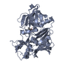 1fknS S: Starting model for refinement |
|---|---|
| Similar structure data |


-Links
 PDBj
PDBj

- Assembly
| Deposited unit | 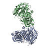
| ||||||||
|---|---|---|---|---|---|---|---|---|---|
| 1 | 
| ||||||||
| 2 | 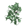
| ||||||||
| Unit cell |
|
-Components
| #1: Protein | Mass: 43627.191 Da / Num. of mol.: 2 / Fragment: Protease Domain Source method: isolated from a genetically manipulated source Source: (gene. exp.) Homo sapiens (human) / Gene: BACE / Production host:  References: UniProt: P56817, Hydrolases; Acting on peptide bonds (peptidases); Aspartic endopeptidases #2: Protein/peptide |   Type: Peptide-like / Class: Inhibitor / Mass: 936.057 Da / Num. of mol.: 2 / Source method: obtained synthetically / Source: (synth.) synthetic construct (others) / References: BETA-SECRETASE INHIBITOR OM00-3 Type: Peptide-like / Class: Inhibitor / Mass: 936.057 Da / Num. of mol.: 2 / Source method: obtained synthetically / Source: (synth.) synthetic construct (others) / References: BETA-SECRETASE INHIBITOR OM00-3#3: Water | ChemComp-HOH / |  Mass: 18.015 Da / Num. of mol.: 450 / Source method: isolated from a natural source / Formula: H2O Mass: 18.015 Da / Num. of mol.: 450 / Source method: isolated from a natural source / Formula: H2OHas protein modification | Y | |
|---|
-Experimental details
-Experiment
| Experiment | Method: X-RAY DIFFRACTION / Number of used crystals: 1 |
|---|
- Sample preparation
| Crystal | Density Matthews: 2.82 Å3/Da / Density % sol: 56.37 % | ||||||||||||||||||||||||
|---|---|---|---|---|---|---|---|---|---|---|---|---|---|---|---|---|---|---|---|---|---|---|---|---|---|
| Crystal grow | Temperature: 293 K / Method: vapor diffusion, hanging drop / pH: 6.5 Details: 22.5% PEG 8000, 0.2 M Ammonium Sulfate, 0.1 M Sodium Cacodylate, pH 6.2, pH 6.5, VAPOR DIFFUSION, HANGING DROP, temperature 293K | ||||||||||||||||||||||||
| Crystal grow | *PLUS Temperature: 20 ℃ / pH: 6.2 | ||||||||||||||||||||||||
| Components of the solutions | *PLUS
|
-Data collection
| Diffraction | Mean temperature: 100 K |
|---|---|
| Diffraction source | Source: ROTATING ANODE / Type: RIGAKU RU300 / Wavelength: 1.5418 Å |
| Detector | Type: MAR scanner 345 mm plate / Detector: IMAGE PLATE / Date: Apr 15, 2001 / Details: mirrors |
| Radiation | Monochromator: multilayer reflector / Protocol: SINGLE WAVELENGTH / Monochromatic (M) / Laue (L): M / Scattering type: x-ray |
| Radiation wavelength | Wavelength: 1.5418 Å / Relative weight: 1 |
| Reflection | Resolution: 2.1→25 Å / Num. all: 59470 / Num. obs: 58812 / % possible obs: 99 % / Redundancy: 3.2 % / Biso Wilson estimate: 23.3 Å2 / Rmerge(I) obs: 0.119 / Net I/σ(I): 7.3 |
| Reflection shell | Resolution: 2.1→2.18 Å / Mean I/σ(I) obs: 2.2 / % possible all: 97.1 |
| Reflection | *PLUS Lowest resolution: 25 Å / Num. obs: 58864 / % possible obs: 98.8 % / Num. measured all: 190727 |
| Reflection shell | *PLUS Highest resolution: 2.1 Å / % possible obs: 97.1 % |
- Processing
| Software |
| ||||||||||||||||||||
|---|---|---|---|---|---|---|---|---|---|---|---|---|---|---|---|---|---|---|---|---|---|
| Refinement | Method to determine structure: MOLECULAR REPLACEMENT Starting model: PDB Entry 1FKN Resolution: 2.1→25 Å / Cross valid method: THROUGHOUT / σ(F): 0 / Stereochemistry target values: Engh & Huber
| ||||||||||||||||||||
| Displacement parameters | Biso mean: 23.1 Å2 | ||||||||||||||||||||
| Refine analyze | Luzzati coordinate error obs: 0.25 Å / Luzzati d res low obs: 5 Å / Luzzati sigma a obs: 0.28 Å | ||||||||||||||||||||
| Refinement step | Cycle: LAST / Resolution: 2.1→25 Å
| ||||||||||||||||||||
| Refine LS restraints |
| ||||||||||||||||||||
| LS refinement shell | Resolution: 2.1→2.23 Å / Rfactor Rfree error: 0.011
| ||||||||||||||||||||
| Refine LS restraints | *PLUS
|
