 Movie
Movie Controller

[English] 日本語
 Yorodumi
- PDB-1lrh: Crystal structure of auxin-binding protein 1 in complex with 1-na... -
+ Open data

- Basic information
| Entry | Database: PDB / ID: 1lrh | |||||||||
|---|---|---|---|---|---|---|---|---|---|---|
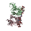
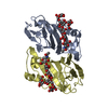
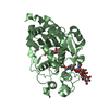
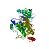
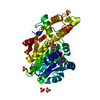
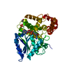
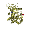
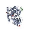
| Title | Crystal structure of auxin-binding protein 1 in complex with 1-naphthalene acetic acid | |||||||||
 Components Components | auxin-binding protein 1 | |||||||||
 Keywords Keywords | PROTEIN BINDING / BETA JELLYROLL / DOUBLE STRANDED PARALLEL BETA HELIX / GERMIN LIKE PROTEIN | |||||||||
| Function / homology |  Function and homology information Function and homology informationpositive regulation of DNA endoreduplication / auxin binding / cytokinesis by cell plate formation / unidimensional cell growth / auxin-activated signaling pathway / positive regulation of cell division / positive regulation of cell size / endoplasmic reticulum lumen / zinc ion binding Similarity search - Function | |||||||||
| Biological species |  | |||||||||
| Method |  X-RAY DIFFRACTION / SYNCHROTRON / difference fourier / Resolution: 1.9 Å X-RAY DIFFRACTION / SYNCHROTRON / difference fourier / Resolution: 1.9 Å | |||||||||
 Authors Authors | Woo, E.J. / Marshall, J. / Bauly, J. / Chen, J.-G. / Venis, M. / Napier, R.M. / Pickersgill, R.W. | |||||||||
 Citation Citation | Journal: EMBO J. / Year: 2002 Title: Crystal structure of auxin-binding protein 1 in complex with auxin. Authors: Woo, E.J. / Marshall, J. / Bauly, J. / Chen, J.G. / Venis, M. / Napier, R.M. / Pickersgill, R.W. | |||||||||
| History |
|
- Structure visualization
| Structure viewer | Molecule: MolmilJmol/JSmol |
|---|
- Downloads & links
-Download
| PDBx/mmCIF format | 1lrh.cif.gz | 153.1 KB | Display | PDBx/mmCIF format |
|---|---|---|---|---|
| PDB format | pdb1lrh.ent.gz | 122.1 KB | Display | PDB format |
| PDBx/mmJSON format | 1lrh.json.gz | Tree view | PDBx/mmJSON format | |
| Others |  Other downloads Other downloads |
-Validation report
| Arichive directory | https://data.pdbj.org/pub/pdb/validation_reports/lr/1lrhftp://data.pdbj.org/pub/pdb/validation_reports/lr/1lrh | HTTPS FTP |
|---|
-Related structure data


-Links
 PDBj
PDBj

- Assembly
| Deposited unit | 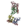
| ||||||||
|---|---|---|---|---|---|---|---|---|---|
| 1 | 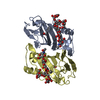
| ||||||||
| 2 | 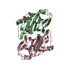
| ||||||||
| Unit cell |
|
-Components
| #1: Protein | Mass: 18411.809 Da / Num. of mol.: 4 / Mutation: D161E/E162Q Source method: isolated from a genetically manipulated source Source: (gene. exp.)  Trichoplusia ni (cabbage looper) / References: UniProt: P13689 Trichoplusia ni (cabbage looper) / References: UniProt: P13689#2: Polysaccharide | alpha-D-mannopyranose-(1-3)-[alpha-D-mannopyranose-(1-6)]alpha-D-mannopyranose-(1-6)-beta-D- ...alpha-D-mannopyranose-(1-3)-[alpha-D-mannopyranose-(1-6)]alpha-D-mannopyranose-(1-6)-beta-D-mannopyranose-(1-4)-2-acetamido-2-deoxy-beta-D-glucopyranose-(1-4)-2-acetamido-2-deoxy-beta-D-glucopyranose Source method: isolated from a genetically manipulated source #3: Chemical | ChemComp-ZN /   Mass: 65.409 Da / Num. of mol.: 4 / Source method: obtained synthetically / Formula: Zn Mass: 65.409 Da / Num. of mol.: 4 / Source method: obtained synthetically / Formula: Zn#4: Chemical | ChemComp-NLA /   Mass: 186.207 Da / Num. of mol.: 4 / Source method: obtained synthetically / Formula: C12H10O2 Mass: 186.207 Da / Num. of mol.: 4 / Source method: obtained synthetically / Formula: C12H10O2#5: Water | ChemComp-HOH / |  Mass: 18.015 Da / Num. of mol.: 375 / Source method: isolated from a natural source / Formula: H2O Mass: 18.015 Da / Num. of mol.: 375 / Source method: isolated from a natural source / Formula: H2OHas protein modification | Y | |
|---|
-Experimental details
-Experiment
| Experiment | Method: X-RAY DIFFRACTION / Number of used crystals: 1 |
|---|
- Sample preparation
| Crystal | Density Matthews: 2.4 Å3/Da / Density % sol: 48.81 % | |||||||||||||||||||||||||||||||||||
|---|---|---|---|---|---|---|---|---|---|---|---|---|---|---|---|---|---|---|---|---|---|---|---|---|---|---|---|---|---|---|---|---|---|---|---|---|
| Crystal grow | Temperature: 298 K / Method: vapor diffusion, hanging drop / pH: 5.5 Details: PEG4000, pH 5.5, VAPOR DIFFUSION, HANGING DROP, temperature 298K | |||||||||||||||||||||||||||||||||||
| Crystal grow | *PLUS Temperature: 291 K / pH: 7 Details: Woo, E.J., (2000) Acta Crystallogr., Sect.D, 56, 1476. | |||||||||||||||||||||||||||||||||||
| Components of the solutions | *PLUS
|
-Data collection
| Diffraction | Mean temperature: 100 K |
|---|---|
| Diffraction source | Source: SYNCHROTRON / Site: SRS  / Beamline: PX9.6 / Wavelength: 0.87 Å / Beamline: PX9.6 / Wavelength: 0.87 Å |
| Detector | Type: ADSC QUANTUM 4 / Detector: CCD |
| Radiation | Protocol: SINGLE WAVELENGTH / Monochromatic (M) / Laue (L): M / Scattering type: x-ray |
| Radiation wavelength | Wavelength: 0.87 Å / Relative weight: 1 |
| Reflection | Resolution: 1.9→30 Å / Num. all: 54476 / Num. obs: 53495 / % possible obs: 98.2 % / Observed criterion σ(F): 0 / Observed criterion σ(I): 0 / Rmerge(I) obs: 0.038 |
| Reflection shell | Resolution: 1.9→1.97 Å / Rmerge(I) obs: 0.096 / % possible all: 90 |
| Reflection | *PLUS % possible obs: 94.2 % |
| Reflection shell | *PLUS % possible obs: 90 % |
- Processing
| Software |
| ||||||||||||||||||||
|---|---|---|---|---|---|---|---|---|---|---|---|---|---|---|---|---|---|---|---|---|---|
| Refinement | Method to determine structure: difference fourier / Resolution: 1.9→15 Å / σ(F): 0 / Stereochemistry target values: Engh & Huber
| ||||||||||||||||||||
| Refinement step | Cycle: LAST / Resolution: 1.9→15 Å
| ||||||||||||||||||||
| Refinement | *PLUS % reflection Rfree: 5 % / Rfactor obs: 0.2 / Rfactor Rfree: 0.24 / Rfactor Rwork: 0.2 | ||||||||||||||||||||
| Solvent computation | *PLUS | ||||||||||||||||||||
| Displacement parameters | *PLUS | ||||||||||||||||||||
| Refine LS restraints | *PLUS
|
